DIFFERENTIAL DIAGNOSIS
Local Adenopathy
Localized lymphadenopathy can suggest the site of infection, although not necessarily the original source of infection. For example, the sexually transmitted disorders usually affect the genitalia and inguinal lymph nodes, but if the primary lesion (e.g., a syphilitic chancre) is located elsewhere, e.g., the mouth, the adenopathy will be in the cervical area. Similarly, diseases spread by insect vectors will cause adenopathy in the area corresponding to the site of inoculation (Table 3).
Table 3: Localized and Generalized Lymphadenopathy
| LOCALIZED LYMPHADENOPATHY | |
| Cervical adenopathy | Pharyngitis
|
Infectious mononucleosis (EBV, CMV)
| |
| Diphtheria (Corynebacterium diphteriae) | |
| Toxoplasmosis | |
| Fusobacterium Infection | |
| Mycobacterial adenitis (M.tuberculosis, M.avium complex etc) | |
| Kikuchi's disease | |
| Kawasaki's disease | |
| Occipital adenopathy | Nonspecific scalp infection |
| Rubella | |
| Peripheral adenopathy | Location varies with inoculation site.
|
Usually axillary or epitrochlear
| |
| Inguinal | Primary syphilis |
| LGV (lymphogranuloma venereum) Chlamydia trachomatis | |
| Chancroid Haemophilus ducreyi | |
| Granuloma inguinale Klebsiella granulomatis | |
| Genital herpes infection, Herpes simplex type 2 | |
| GENERAL LYMPHADENOPATHY | |
| |
Cervical Adenitis
Cervical adenitis is usually secondary to pharyngitis by respiratory viruses or by group A streptococcus. Group A streptococcal infection (Streptococcus pyogenes) of the pharynx can cause tender anterior cervical adenopathy. The appearance of the pharynx, the presence of exudative material, and the character of the nodes are all nonspecific.
Epstein Barr Virus:
Infectious mononucleosis is usually associated with a pharyngitis that may be exudative. Cervical adenitis involving both the anterior and posterior cervical nodes is almost always present. Mononucleosis should be considered as a cause of pharyngitis in the adolescent or young adult. The lymphadenopathy may not be localized to just the cervical region but may be generalized. Splenomegaly, palatal petechiae, supraorbital edema and a generalized maculopapular rash may occur. Hepatomegaly and/or abnormalities of liver function are common in infectious mononucleosis.
Cytomegalovirus:
Cytomegalovirus (CMV) infection can mimic infectious mononucleosis, but the Monospot and EBV antibody tests are negative. The pharyngitis and cervical adenopathy in CMV infection is less pronounced than in EBV mononucleosis.
Rarer bacterial etiologies of pharyngitis include Neisseria gonorrhoae (severe pain but minimal erythema), A. hemolyticum (may be accompanied by a diffuse erythema) and Yersinia species (sometimes suggested by gastrointestinal symptomatology).
Mycobacterial Species:Corynebacterium Diptheriae:Scrofula (tuberculous cervical lymphadenitis) presents as painless, enlarged and matted lymphnodes; fluctuation may develop in some nodes. Spontaneous drainage of caseous material onto the skin surface (scrofuloderma) may occur. The node enlargement is usually unilateral and isolated to one group of nodes. The disease is usually located to the lymph nodes only. This syndrome is typically seen in children who are otherwise healthy and have a negative chest x-ray. The responsible pathogens are M. scrofulaceum and M. avium complex (MAC). Mycobacterial cervical adenitis in young adults, aged 20 to 40, is usually caused by classical M. tuberculosis.
Vietnamese man with tuberculosis lymphadenitis
Diphtheria is an extremely rare cause of acute cervical lymphadenopathy except in developing countries. History of immunization is crucial. Diphtheria should be suspected in any nonimmunized patient with a pharyngitis accompanied by a gray pseudomembrane that is difficult to remove. Cervical adenopathy and edema of the anterior neck and submandibular areas frequently occur with this form of pharyngitis. Other much more frequent causes of a pharyngeal pseudomembrane are EBV, Streptococcus group A, fusobacterium, Candida albicans, and upper respiratory tract viral pathogens.Kawasaki’s Disease:
The mucocutaneous lymph node syndrome (MLNS) is characterized by protracted fever (longer than five days) associated with cervical adenopathy, conjunctival injection, redness of pharynx and lips, and diffuse scarlatiniform rash including the palms and soles, with subsequent desquamation. The disease may be mistaken for Group A streptococcal scarlet fever, but the diagnostic studies for streptococcal infection are negative and penicillin is ineffective. The fatality rate (about 1%) is related to coronary arteritis.Kikuchi’s Disease:
Kikuchi’s disease (also called Kikuchi-Fujimoto disease or Kikuchi’s histiocytic necrotizing lymphadenitis) occurs predominately in females. Clinical features are characterized by fever, cervical lymphadenopathy, and transient skin rash. Signs and symptoms usually resolve spontaneously within one to four months. The definitive diagnosis of Kikuchi’s disease is made by lymph node biopsy. Histological findings show follicular hyperplasia in early phase, the presence of blast cells in the “proliferative phase”, and necrosis with histiocytes in the “necrotizing phase”. This syndrome is more common in Asia than elsewhere.Other Nonbacterial Causes:
Adenovirus and herpes simplex type 1 can produce severe exudative and ulcerative pharyngitis, as can primary HIV infection. Mycoplasma may cause a mild pharyngitis similar to viral infection. Cervical adenopathy may be mimicked by other inflammatory processes of the neck including parotitis and Ludwig’s angina.
Occipital Lymphadenopathy
Local infections of the scalp such as pediculosis capitis, impetigo, and ringworm may cause lymphadenopathy in the occipital region. Systemic infections such as syphilis and tuberculosis may enlarge occipital nodes, but usually involve other nodes more dramatically. Rubella virus causes generalized infection that involves occipital nodes; this adenopathy may precede the rubella rash by several days. The nodes, although generalized, usually predominate in occipital, mastoid, and posterior cervical locations. Rubeola also can cause this pattern of adenopathy in addition to rash, fever, and upper respiratory symptoms.
Peripheral Axillary and Epitrochlear Adenopathy
Enlargement of the axillary and epitrochlear or inguinal nodes occurs most frequently from bacterial infection on the extremities. Repeated trauma to the lower extremities can lead to inguinal lymphadenopathy. Nonspecific inguinal adenopathy is common such that biopsy of the inguinal nodes is usually nondiagnostic. If other nodes are enlarged and accessible, biopsy may be more productive there.
Group A Streptococcus:
Group A streptococcal infection of the extremities may produce cellulitis and lymphangitis with regional adenopathy. Fever and superficial red streaks may occur, with painful and tender swollen regional nodes.
Francisella Tularensis:
Tularemia of the ulceroglandular type is most frequently associated with the bite of a tick or deerfly, or with contact with the carcass of an infected animal. The site of inoculation is characterized by an ulcerating papule. Regional adenopathy, the location of which depends on the inoculation site, is manifested by hot, large, fluctuant, and tender lymph nodes that may suppurate and drain spontaneously. On the other hand, the node enlargement may at times be much less dramatic and recognized only after very careful physical examination.
Bartonella Species:
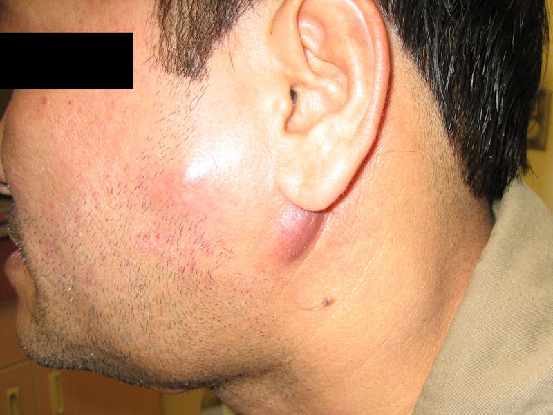
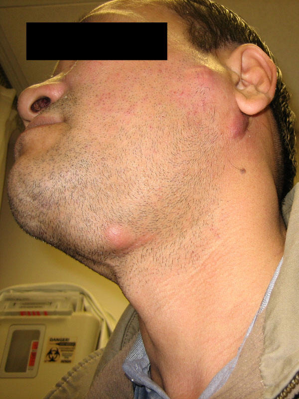
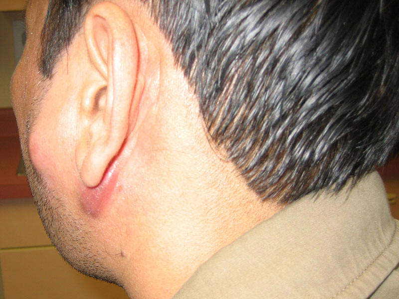
Cat Scratch Disease may be manifest by localized adenitis in the epitrochlear, axillary, and inguinal areas depending on the site of the scratch or bite. The etiology of cat-scratch disease is Bartonella henselae, a fastidious gram-negative rod. Facial lesions may be associated with cervical or auricular adenopathy. The primary lesion is a tender papule that is frequently capped by a vesicle. Regional nodes then become enlarged and tender, and the overlying skin may be red. If the node becomes fluctuant, it should be aspirated but not incised. Aspiration may reduce local pain with a dramatic lysis of fever.
Yersinia Pestis:
The most common form of disease, bubonic plague, is manifested by high fever and systemic toxicity, as well as by an erythematous, edematous matted group of inguinal or axillary nodes that can suppurate and drain spontaneously. The inoculating flea bite is usually not apparent. The enlarged lymph node, the bubo, is extremely tender. The diagnosis can be made by culture of an aspirated lymph node.
Spirillium Minus:
Rat-bite fever caused by S. minus may cause rash, lead to the syndrome of a swelling at the site of the bite, regional adenopathy, and fever. The local infected nodes are tender, firm, and freely movable. One to four weeks after the bite, the local lesion flares again as the systemic phase of the illness begins. Fever, generalized rash, and arthritis occur. This infection is more common in Asia and is called Sodoku in Japan. In the other form of rat-bite fever, which is more common in the Western hemisphere is caused by Streptobacillus moniliformis, adenopathy is not prominent.
Sporotrichum Schenkii:
Sporotrichosis is often manifest by primary lesion of the upper extremities and the characteristic intermittent hard red lumps along the lymphatics. The nodes then enlarge and the cutaneous nodules may ulcerate. Pain and fever are absent, and the streaking seen in acute bacterial lymphangitis is rare. Sporotrichosis is diagnosed by culture of the nodules, since neither serology nor scrapings or the lesions for staining are helpful. Usually the infection stays localized but requires a long time for resolution. Although rare, primary cutaneous coccidioidomycosis, nocardiosis, and blastomycosis may mimic local sporotrichosis.
Herpes Zoster:
Most commonly seen on the trunk, herpes zoster causes a syndrome of clustered vesicles in a unilateral segmental distribution, frequently preceded and accompanied by marked paresthesias, mild fever, and headache. Signs and symptoms usually precede the rash by a few days but at times by as long as one to two weeks. Regional adenitis is a consistent feature of herpes zoster. The nodes are nontender and freely movable.
Herpes Simplex (Herpetic whitlow):
Epitrochlear and axillary adenopathy may be a complication of herpetic whitlow, an infection of the pulp of a finger caused by Herpes simplex, usually type 1. This primary lesion is usually very painful.
Rickettsioses:
Rickettsial infection present with fever, headache, myalgic and regional lymphadenopathy. The etiologies include Oritentia tsutsugamushi (Scrub Typhus), Rickettsia akari (Rickettsialpox), and other spotted fever group Rickettsial infections including R. australis (Queensland tick typhus), R. honei (Flinders Island spotted fever), R. conorii (African tick bite fever), R. sibirica (Siberian tick typhus), R. sibirica mongolotimonae (Lymphangitis-associated rickettsiosis), R. slovaca (Tick-borne lymphadenopathy).
Inguinal Adenopathy
Many sexually transmitted infections are responsible for inguinal lymph node enlargement. Frequently, a genital ulcer accompanies the adenopathy, producing the “ulcer-adenopathy” syndrome.
Treponema Pallidum:
Syphilis often presents as a painless skin lesion, the chancre, usually on the external genitalia, with enlarged freely movable, firm, nonsuppurating, and painless inguinal nodes. The chancre heals in two to six weeks, but the lymphadenopathy may persist for months. Diagnosis of primary syphilis is made by a characteristic lesion and serology.
Haemophilus Ducreyi:
Unlike syphilis, the genital ulcers and inguinal nodes of chancroid are tender and painful. If untreated, the nodes may suppurate and rupture to form a large single ulcer. This last complication may be prevented by aspiration when the node is tense.
Lymphogranuloma Venereum (LGV):
Chylamydia trachomatis is the cause of Lymphogranuloma vereum, a genital ulcer disease. Inguinal adenopathy is prominent in LGV and may progress to multiocular suppuration with multiple fistulae. At times the inguinal ligament forms a line of cleavage through the matted nodes, which are enlarged both above and below the ligament (the “Groove” sign). The primary genital lesion is a single small ulceration that generally goes unnoticed. It has usually disappeared by the time the lymph node enlargement occurs. LGV may be accompanied by generalized constitutional symptoms and is occasionally associated with severe arthralgias, diarrhea, and hyperglobulinemia. It is rare in developing countries but can be endemic in tropical countries. PCR of specimens taken from the ulcer is now the most sensitive diagnostic method.
Klebsiella Granulomatosis:
Klebsiella granulomatous is the cause of granuloma inguinale (Donovanosis). The disease is manifested by a painless genital papule anad nodule that erodes to leave a beefy red granular base. The infection spreads to the inguinal area and produces a marked subcutaneous swelling. The lesion is called a “pseudobubo” since it is not really due to lymph node involvement, but rather to soft tissue granulation and frequently ulceration. This infection is rare in developing countries.
Herpes Viruses:
Genital herpetic lesions are characteristically multiple, small erosions on an erythematous base, typically arranged in a cluster. They are extremely painful and are often accompanied by bilateral or unilateral inguinal adenopathy.
Generalized Adenopathy
Fever and generalized adenopathy are usually due to viruses, especially EBV and rubella, and HIV. EBV mononucleosis may also be mimicked by infection with CMV, Toxoplasmosis gondii and HIV. Acquired or reactivated CMV infection is most frequent in normal young people, in patients who have had surgery with bypass (postperfusion syndrome), recipients of blood transfusions, leukocyte infusions and transplants, and in immunocompromised individuals. Patients with this CMV syndrome may present with protracted fever and with moderate hepatic inflammation along with lymphocytosis and atypical lymphocytes, which occur late in the course of the disease. Splenomegaly and adenopathy are not always present. The hepatitis viruses and HHV-6, the cause of roseola infantium, can also produce lymphadenopathy and a mononucleosis-like illness. Other viruses, particularly rubella, adenovirus, and dengue can also cause significant generalized lymph node enlargement (Table 3).
Primary HIV infection causes a mononucleosis-like illness often accompanied by a diffuse salmon-colored rash that includes the palms and soles. Lymphadenopathy occurs in 50 to 75 % of patients who develop an acute illness approximately 3 to 6 weeks after initial exposure to HIV. In the latency period, persistent generalized lymphadenopathy (PGL) affects between 50 to 70 percent of HIV patients. The lymph nodes are non tender and affect the nodes in the cervical, mandible, inquinal and axillary areas.
Lynphadenopathy is found in 5% of acute viral hepatitis. HBV infection in childhood can be associated with papular dermatitis with enlarged lymph nodes in the axillary and inguinal regions-the Gianotti-Crosti syndrome. This syndrome can also occur after infection by EBV, HAV, HCV, CMV, enterovirus, parvovirus, parainfluenzavirus, rotavirus, respiratory syncytial virus, HIV, HHV-6, Mycoplasma pneumoniae, beta-hemolytic streptococci, Bartonella henselae, Borrelia burgdorferi, and following vaccination with influenza virus vaccine, measles-mumps-rubella vaccine, hepatitis B vaccine, oral polio vaccine or Japanese encephalitis vaccine.
Toxoplasma gondii:
The most common syndrome seen in acquired symptomatic toxoplasmosis resembles EBV mononucleosis, with enlarged nonsuppurative lymph nodes, fever, and maculopapular rash. The nodes in toxoplasmosis are firm, discreet, freely movable, and at times, painful and tender. The disease has occasionally been misdiagnosed as Hodgkin’s disease. Frequently the cervical nodes are the largest, increasing the clinical similarity of this entity to mononucleosis. Toxoplasmosis can involve the CNS, liver, heart, lungs, or eyes as target organs. This infection must be considered in patients with an underlying immunosuppressive disease.
Brucella Species:
Brucellosis is characterized by fever, chills, hepatosplenomegaly, arthralgia, and leukopenia with relative lymphocytosis. Sarcoiliitis and spondylitis are common. Small, nontender, cervical and axillary node enlargements are common. These nodes may show granulomas or a striking reticuloendothelial hyperplasia with some multinucleate giant cells. Testicular swelling and/or pain may occur.
Histoplasmosis and fungi:
Disseminated histoplasmosis and other deep fungal infections are capable of producing widespread lymphadenopathy as part of the generalized reticuloendothelial reaction to these agents.
Treponema pallidum:
Secondary syphilis is recognized by nontender, generalized lymphadenopathy with fever and a diffuse nonpruritic maculopapular rash, which often includes the palms and soles. The nodes may remain palpable even months after the disappearance of other stigmata of secondary syphilis, i.e., fever, rash, condylomata, and mucous patches. Pre and postauricular, occipital and epitrochlear nodes are all frequently enlarged in secondary syphilis.
Mycobacterium tuberculosis:
Non-Infectious CausesMiliary tuberculosis may cause generalized adenopathy. The typical military pattern on chest x-ray may often be delayed.
Many noninfectious disorders include adenopathy among their manifestations, e.g., lymphoreticular malignancies, collagen vascular diseases, and sarcoidosis (Table 2). Peripheral non-tender, lymphadenopathy is common in sarcoidosis; the cervical, axillary, epitrochlear, and inguinal nodes are the most commonly involved regions. The nodes are non-ulcerative unlike infectious causes.
Table 2: Infections Causing Lymphadenopathy
Infections Viral
- Infectious mononucleosis (Epstein-Barr virus)
- Infectious hepatitis
- Adenovirus
- Herpes simplex, Herpes zoster
- HHV-6 (Human herpesvirus-6)
- HIV
- Human T-lymphotropic virus
- Rubella
- Measles
Bacterial
- Cutaneous infections (Staphylococcus, Streptococcus)
- Cat-scratch fever
- Chancroid
- Melioidosis
- Tuberculosis
- Atypical Mycobacteria
- Primary and secondary syphilis
Chlamydial
- Lymphogranuloma venereum
Protozoan
- Toxoplasmosis
Mycotic
- Histoplasmosis
- Coccidiodomycosis
Rickettial
- Scrub typhus
Helminthic
- Filariasis
Noninfectious Causes Autoimmune
- Rheumatoid arthritis
- Juvenile rheumatoid arthritis
- Systemic lupus erythematosus
- Dermatomyositis
- Mixed connective tissue disease
- Sjogren’s syndrome
Iatrogenic hypersensitivity
- Serum sickness
- Drug hypersensitivity
- Anticonvulsants Phenytoin, carbamazepine, primidone
- Antibiotics Sulfas, penicillins, cephalosporines, erythromycin
- Anti-inflammatory Gold, sulindac, sulfasalazine
Neoplasm
- Lymphoreticular malignancies
- Paraproteinemias (monoclonal gammopathy)
- Metastatic diseases
Miscellaneous
- Sarcoidosis
- Amyloidosis
- Vaccine-induced
- Kikuchi's disease
- Hyperthyroidism
- Storage diseases (Gaucher, Niemann-Pick, Fabry, and Tangier)
- Severe hypertriglyceridemia
- Kimura’s disease
- Kawasaki disease
- Silicone injection
- Graft vs host disease
- Autoimmune hemolytic anemia
- Gianotti-Crosti syndrome
No comments:
Post a Comment