Your Candida Albicans Symptoms Can Be Misdiagnosed For Years
Candida
albicans symptoms indicate the presence of an organism which is as
dangerous as it is difficult to diagnose. Thank God there are
home remedies which are effective against it.The challenge is to recognize the symptoms.
Candida
albicans symptoms are so diverse and are involved in so many different
conditions that diagnosis is very difficult. Candida symptoms are not
only diverse but are seemingly unrelated too.
It is for this
reason that you may go from doctor to doctor who will often tell you
with a straight face, that nothing is REALLY wrong with you, and that
it is all in your mind.
It is for this reason that you may experience
chronic yeast infection and yet the doctors will continue to prescribe topical anti-fungal treatment, which does not address the REAL problem
To
add to the confusion, many persons, including doctors, seem to think
that candida albicans largely manifests in relatively minor health
conditions such as vaginal yeast infections or oral thrush, which can
easily be dealt with, they advise, with a treatment of anti-fungal
medications.
While candida albicans can cause yeast infections
and oral thrush, its existence in its pathogenic or disease-causing
form (or hyphal form) and the widespread effects of its overgrowth is
not given much attention.

Candida Albicans Symptoms- The Real Story Behind It All
Candida
albicans is a fungus which can exist as yeast or as an organism which
has long branches called hyphae, by which it feeds and extends its
growth.Candida albicans is one form of yeast, and it resides quite
harmlessly on our bodies and in our intestines. The harmless existence
of candida is ensured by the proper functioning of your immune system
and the correct balance of yeast and other microorganisms within your GI
(gastrointestinal) tract.
If conditions cause a disruption of
the balance in such a way that your “friendly “ bacteria are
overwhelmed by other bacteria, or all intestinal flora are killed off,
yeast takes the opportunity into change to its pathogenic, or
disease-causing form and grows its hyphae.
This can occur for
example, after prolonged antibiotic use. Antibiotics destroy not only
unfriendly bacteria, but also friendly bacteria. In this newly created
space, and environment,candida albicans will opportunistically
overgrow.
Return to
the top of this page
Birth
control pills, too much sugar in the diet and a compromised immune
system are also factors which can alter your internal environment.
In
the hyphal form candida produces toxins into your blood stream which
can affect the functioning of every organ and system in your body.
In
addition, candida can permeate your intestinal walls and thus cause
undigested food, bacteria and toxins to enter your bloodstream. Your
immune system tries to deal with these foreign bodies and soon you are
suffering from food allergies and other inflammatory responses wherever
the foreign bodies may be located.
It has been determined that Candida produces as many as 79 different toxins.
As
if that is not bad enough, the candida fungus can enter your blood
stream and make its home in any place in your body. After a while your
entire body can be overloaded with not only the yeast itself but also
the toxins from it.
Yeast can also overgrow in warm surfaces on
your body such as in your mouth, the genital areas, under the breasts.
Treating the outbreak at the site is not a permanent solution as the
fungus will continue to return. The answer is to seek to maintain the
bodily pH at a level which discourages the overgrowth of candida and
bring about
candida albicans symptoms.
Return to
the top of this page
Candida Albicans Symptoms
Given
the nature of Candida, how can you even begin to think that problems
which occur in your body may be candida albicans symptoms?A good answer
is that the very fact that you are experiencing symptoms which are
unrelated may be, in and of itself, a Candida albicans symptom.
And
if tests return "Normal" and you KNOW you are not 100% well, this is
another red flag. A test for an overgrowth is inconclusive as candida
albicans is normally resident in your body, and it is hard to tell
there is an overgrowth from clinical tests alone.
In order for
you to make some sense of the diversity, let us organize the symptoms
(and we cannot name all of them) according to the functions they affect
or where they occur, knowing that it may be just an indication of
wider systemic disorder.
The main areas of candida albicans activity are:
• The Gastrointestinal Tract (or GI Tract)
• The Genito–urinary Tract and Reproductive System
• Emotional and Hormonal /Nervous System
• The Respiratory System
As
you peruse the following symptoms, try to recall when your symptoms
occurred and how you felt as no one could say conclusively, why it was
happening.
Some have been given tranquilizers as their doctors felt they may be neurotic.
Return to
the top of this page
Symptoms In The Gastrointestinal Tract
In
the gastrointestinal tract or digestive tract (which extends from the
mouth to the anus) is one area where you will experience candida
albicans symptoms. Common symptoms include:
• Belching, bloating, acid reflux, Nausea, indigestion, gastritis
• Diarrhea, gas, colitis, Food allergies and food cravings and abdominal pain
• Bad breath, white coating on your tongue, mouth blisters and bleeding gums
Symptoms in The Genitourinary Tract And Reproductive System
Candida
albicans symptoms in the genito-urinary tract (includes all the
muscles and organs which produce, store and remove urine) and the
reproductive system:• Painful intercourse, burning during urination,
impotency, itching of the genitals and rectum
• Infertility, irregular menstruation, vaginal yeast infection, premenstrual syndrome(PMS)
• Redness and soreness of the vulva, vaginal discharge, kidney and bladder infections
Symptoms of An Emotional Or Mental Nature
You
may experience candida albicans symptoms that are of an emotional or
mental nature such as:• Anxiety, confusion, depression, foggy thinking,
panic, irritability, apathy
• Bumping into things, frequent headaches, mood swings, memory problems,
• Unreasonable anger, dizzy feelings, loss of concentration, autism
Return to
the top of this page
Symptoms in Your Respiratory System
Candida
can also affect your respiratory system with the following candida
albicans symptoms:• Sneezing, wheezing, congestion, coughing, shortness
of breath
• Bronchitis, Asthma, Repeated bouts of sinus infection, Pneumonia
CAndida Albicans Symptoms On Your Skin
Your
skin is another organ which may be subject to candida albicans symptoms
which include:• Athlete’s Foot, fingernail and toenail fungus, skin
rashes, scaly skin
• Scaly scalp, Diaper rash, eczema
Candida Albicans And Disorders It May Be Associated With
Candida
albicans tends to permeate the membranes of your tissues. Your immune
system has to also deal with the toxins produced and everyday attacks
from your environment.Eventually your immune system is overwhelmed and
pathogens will be going after different organs in your body. Had it
not been for the overload caused by candida, your immune system would
have been able to withstand the other attacks.
The situation can
become life-threatening as it can cause septic shock. It is in this way
that candida can be associated in the development of the following
(and

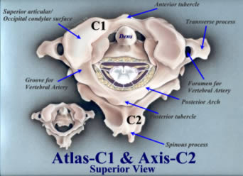
 Figure
1: Upper Cervical Spine – No subluxation
Figure
1: Upper Cervical Spine – No subluxation Figure
2: Upper Cervical Spine – Skull to atlas subluxation
Figure
2: Upper Cervical Spine – Skull to atlas subluxation 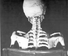 Figure
3: Even Death does not hide the proof!
Figure
3: Even Death does not hide the proof! 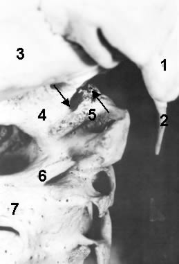 Figure
4: A close look at the subluxation!
Figure
4: A close look at the subluxation!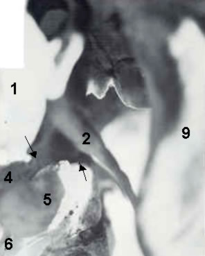 Figure
5: An even closer look at the subluxation!
Figure
5: An even closer look at the subluxation!  Figure
6: The Effects of the Atlas Subluxation
Figure
6: The Effects of the Atlas Subluxation Figure
7: The Effects of the Atlas Subluxation
Figure
7: The Effects of the Atlas Subluxation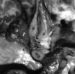 Figure 8: Myodural Bridge
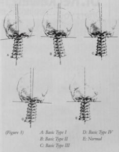


 |
| 
 There has been a new food revolution in the past two decades which
has changed the way we eat forever. Health-conscious farmers have begun
to turn away from traditional corn- and grain-based diets for cows and
let them eat grass in pastures, just as nature originally intended.
There has been a new food revolution in the past two decades which
has changed the way we eat forever. Health-conscious farmers have begun
to turn away from traditional corn- and grain-based diets for cows and
let them eat grass in pastures, just as nature originally intended.