Tuesday, 21 February 2012
bladder meridian causes spasm
before I go for a wee there is a tight feeling in my right leg, that tightness subsides as soon as I start to wee
Monday, 20 February 2012
Thisisms JIMMYlEGS my reply to warning- my info
I suppose I thought if this woman is going to ban me for nothing, then I will give her good reason. What does concern me about this site, is they have access to anyone from any country as the have a language interpreting system so any language can be converted into English, thats what worries me the range of people that they can affect. I am sure that the good people that originally set up this site 10 years ago with good intentions and built up a good strong ms community are not in charge of it now. I think it is now in the wrong hands.Oh and I say woman not to be disrespectful, but apparently he is a she, possibly meaning that she is not the original Jimmylegs.
JIMMYLEGS
Ban me for telling the truth, ask me if I care, because Madame "what goes around comes around" I think bottom line for you all. The professor included "The game is up" and I am not the only one who's clocked it, "so time will tell" the scam in duping new and vulnerable visitor. befriending them and adorning them with all your kind vit advice, sympathy heart and flowers, Then DougL sterring them in the Ccsvi direction is wrong manipulative, and CECE bumping up ccsvi topics on a constant basis must be your dream.
So excuse me if I am not quaking in my boots, and crying ooooh! I'm scared, then you dont know me at all, do your worst, fill your boots. It 'aint me that should be worried
You banned Fee001, Fee002 Goodtotalk, Sunnee go the whole hog ban Icecube, it was an in joke anyway to come back as someone who is chilled and cool, I'm even smiling now.
So thanx 4 making me so jolly today, I'll keep watching and come back on a different computer sysrem with yet another name, but next time I wont give you N E clues as to whome I am.
So enjoy! while it lasts
Your/regards
Fiona
Friday, 17 February 2012
Thisisms- powerful words but not true
Cheer and everyone else
These are exact words used to advertise this forum, why should this statement be declared, to gain new vulnerable and possibly very young and most importantly impressionable people to a forum which has only one directive.
In UK a 14 year old with a user name of UAE4EVA posted on another forum, she has been diagnosed with ms, which surely must be frightening, thrust into a world of uncertainty for a young girl only a year older than my own son.
The cause of CCSVI is unproven, I and many others believe that there is possibly another reason why it occurs, others deserve to know that, in not allowing that to happen, by actively deleting or driving others away who have that alternative opinion is manipulative and very very wrong to do so.
And that is my point.and my last statement on the matter.
......................................................................................................................................................................
News News of ThisIsMS
Site map of ThisIsMS » Forum : ThisIsMS
Welcome to the world's leading forum on Multiple Sclerosis research, support, and knowledge. For over 10 years, This is MS has provided an unbiased community dedicated to Multiple Sclerosis patients, caregivers, and affected loved ones.
Thisisms site yet again distracts me. as this statement is untrue
 News of ThisIsMS
Site map of ThisIsMS » Forum : ThisIsMS
Welcome to the world's leading forum on Multiple Sclerosis research, support, and knowledge. For over 10 years, This is MS has provided an unbiased community dedicated to Multiple Sclerosis patients, caregivers, and affected loved ones.UAE4EVA I think of her nearly every day, just 14
my attention got diverted by other issues. but now my focus is back on. When i start to feel sorry for myself, I think of UAE4EVA and realize, that I have no right
For all UAE4EVAs out there
This young girl aged just 14 has been diagnosed with ms.this will be my last post for this year, because the most powerful post. on this blog
The life of this young girl and that of her parents were DESTROYED at one moment with one little sentence "you have ms" my son is 13 a year younger, just beginning his life as a teenager does, A NEW him, new ideas, new personality, NEW LIFE. what has been done to this girl is unforgivable, her life has been snuffed out, written off, by mistaken arrogant, men in suits, who have had the attitude that the only ALTERNATIVE is by finding a CURE. A cure for something that has been fabricated and made up over the years, with the help of gossip and assumption which has been able to grow under the nose of the MS SOCIETY an organization which is supposed to help. RUBBISH!!!!!
I cant tell this young girl that they have got it WRONG, I cant tell her parents, as I dont know who they are either, and she is not now present on the new ms society boards, as she is under the age to be on them, and that is in a way a very good thing.
I know I am FIXABLE and am doing that, but she does not, her Christmas will be one of isolation and sadness. HOW DARE THEY!!! do this to her.
I will continue to FIGHT and be HEARD for all the UAE4EVAs out there, and that my friends is a lot of people.
What they have done and continue to do so in ignorance is SHOCKING, and although I am feeling very good and happy, this truly BREAKS MY HEART to watch.
Thursday, 16 February 2012
Parkinson, Minieres syndrrome, and trigeminal Neuralgia
Parkinson's Disease, Meniere's Syndrome, Trigeminal Neuralgia and Bell's Palsy: One Cause, One Correction
By Michael T. Burcon, DC
Abstract I currently have 16 Meniere's syndrome, two Parkinson's disease, two Trigeminal neuralgia and two Bell's palsy patients under my care. They all have one thing in common: The atlas vertebra is subluxated posteriorly, which has caused the head to project forward, taking away the healthy curve of the neck. In each patient, the pelvis has twisted to take pressure off the important nerves in the upper neck and brainstem, causing one leg to appear shorter than the other; normal lumbar curvature is compromised; and finally, if not specifically adjusted, the patient compensates by developing an exaggerated curve in the thoracic spine.
I hypothesize that in each patient, kink(s) in the neck inhibited the normal flow of cerebrospinal fluid out of the skull and down the spine; this created excess pressure in the fourth ventricle, causing abnormal function of some of the structures in the midbrain. It also inhibited the flow of blood into the occipital area of the brain by kinking one of the vertebral arteries. Additionally, I suggest that the posterior atlas irritated the anterolateral aspect of the brainstem, irritating any combination of the bottom seven cranial nerves.
All 22 patients improved dramatically after one or two adjustments under cervical-specific chiropractic care. When the atlas returns to juxtaposition, the spinal cord relaxes and actually positions itself lower within the spinal column.
Key Terms: Parkinson's disease; Meniere's syndrome; Trigeminal neuralgia; Bell's palsy, posterior atlas subluxation; specific adjustment.
Introduction
Parkinson's disease (PD, Paralysis Agitans, or "Shaking Palsy") is an idiopathic, slowly progressive, degenerative central nervous system (CNS) disorder with four characteristic features: slowness and poverty of movement; muscular rigidity; resting tremor; and postural instability. Parkinson's disease is the fourth-most-common neurodegenerative disease afflicting the elderly: It affects about 1 percent of the population over 65 years old, compared with 0.4 percent of the population under 40 years old. The mean age of onset is about 57 years of age. Onset in childhood or adolescence (juvenile Parkinsonism) also occurs.1
The etiology and pathophysiology of primary Parkinsonism is loss of the pigmented neurons of the substantia nigra, locus ceruleus and other brainstem dopaminergic cell groups. The loss of substantia nigra neurons, which project to the caudate nucleus and putamen, results in depletion of the neurotransmitter dopamine in these areas.1
For 50 percent to 80 percent of patients with PD, the disease begins insidiously with a resting 4- to 8-Hz "pill-rolling" tremor of one hand. The tremor is maximal at rest; diminishes during movement; is absent during sleep; and is enhanced by emotional tension or fatigue. The hands, arms and legs usually are most affected, in that order. The jaw, tongue, forehead and eyelids also may be involved, although the voice is not affected. Many patients display only rigidity and never manifest tremor. Progressive rigidity, slowness and poverty of movement (bradykinesia) and difficulty in initiating movement (akinesia) follow.1
The face becomes mask-like and open-mouthed, with diminished blinking. Posture becomes stooped. Patients find it difficult to start walking; the gait becomes a shuffle with short steps and the arms are held flexed to the waist and fail to wing with stride. The steps may inadvertently quicken, and the patient may break into a run to keep from falling ("festination"). On examination, passive movement of the limbs is met with plastic, unvarying lead-pipe rigidity; superimposed tremor bursts may give ratchet-like cogwheel quality.1
The sensory examination usually is normal. Signs of autonomic nervous system function may be seen. Muscle strength usually is normal. Dementia occurs in about 50 percent of patients; depression also is common.1
The standard medical treatment for PD has been the administration of the drug Sinemet, which combines Levodopa (a short-acting drug that enters the brain and is converted into dopamine) and Carbidopa (which enhances Levodopa's action in the brain). Several neurosurgical techniques also exist, including thalamotomy (destruction of the ventral thalamus to control tremor); pallidotomy (destruction of the posterior ventral globus pallidus to control hyperkinetic symptoms); and deep- brain stimulation (electrode implantation for patient-controlled stimulation of the thalamus to control tremor). While medication and surgery may control symptoms temporarily, neither stops or reverses the progressive degeneration of the substantia nigra.2
B.J. Palmer reported the use of upper-cervical chiropractic care with PD patients as early as 1934. In his writings, he referred to patients with shaking palsy and listed improvement or correction of symptoms such as tremor; shaking; muscle cramps and/or contracture; joint stiffness; fatigue; lack of coordination; difficulty walking, or inability to walk; numbness; pain; and muscle weakness. His chiropractic care included paraspinal thermal scanning using a neurocalometer (NCM); a cervical radiographic series to analyze the upper-cervical spine; and a specific upper-cervical adjustment performed by hand. Erin L. Elster, DC, found no other references for the chiropractic management of PD patients, prior to her study on 10 PD patients in the year 2000, utilizing modern upper-cervical chiropractic care.2
Three-month re-evaluations revealed substantial improvement in subjective and objective findings in six of the 10 patients, and mild improvement in two patients. The findings of the other two patients, both over age 65, remained unchanged. According to the Unified Parkinson's Disease Rating Scale (UPDRS), six of 10 patients showed overall improvement ranging from 21 percent to 43 percent after three months of upper-cervical chiropractic care.2
Meniere's syndrome is characterized by vertigo or dizziness, and some combination of four associated symptoms: nausea, inner-ear pressure, low-frequency hearing loss and tinnitus. The cause of Meniere's syndrome is unknown and the pathology is poorly understood.1 The attacks of vertigo appear suddenly, last from a few to 24 hours, then subside gradually. The attacks are associated with nausea and vomiting. The patient may feel a recurrent feeling of fullness in the affected ear, and the hearing in that ear tends to fluctuate, but worsens over the years. Tinnitus may be constant or intermittent.
Trigeminal neuralgia (Tic Douloureux) is a disorder of the trigeminal nerve producing bouts of severe, lancinating pain lasting seconds to minutes in the distribution of one or more of its sensory divisions, most often the mandibular and/or maxillary. The cause is uncertain. Recently, surgery at autopsy suggests that this condition is essentially a compressive neuropathy of the brainstem.1
Bell's palsy is a unilateral facial paralysis of sudden onset and unknown cause. Pain behind the ear may precede the facial weakness that develops within hours, sometimes to complete paralysis. The mechanism is presumed to involve swelling and compression of the facial nerve. (1)
In addition to the upper-cervical chiropractic care based on the research of B.J. Palmer, with the assistance of Lyle Sherman, DC (later refined by William G. Blair, DC), I have added the lower-cervical research and adjustment utilized by Walter Vern Pierce, DC, into a technique that I refer to as "cervical-specific chiropractic."3
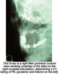
In my previous research with cases involving brainstem irritation (Meniere's disease, Trigeminal neuralgia and Bell's palsy), I discovered that the main cause was cervical trauma. The trauma forced the top cervical vertebra (atlas) to subluxate posteriorly, with laterality on the opposite side of the patient's symptoms (i.e., if the patient had fullness and gradual loss of hearing in the right ear, the atlas listing would be posterior and inferior on the left [PIL]). These same findings are substantiated by my Parkinson's research.4
Methods
My technique is based on the work of B.J. Palmer, as developed at his research clinic at Palmer Chiropractic College in Davenport, Iowa, from the early 1930s until his death in 1961.5-7 I have also studied the vertebral subluxation pattern work of B.J.'s clinic director, Lyle Sherman, DC, for whom Sherman College of Straight Chiropractic, is named.8
A detailed case history is taken on the first visit, followed by a spinal examination. First, the patient's cervical spine is graphed, using an advancement of the dual-probed NCM first used by B.J.9 Next, cervical motion palpation is performed, noting any aberrant motion of the vertebrae.
Detailed leg checks are performed on each patient visit, utilizing the work of J. Clay Thompson, DC, and Clarence Prill, DC.10 With the patient prone, an apparent short leg often is noted. The patient is instructed to turn his or her head to the right. If the short leg becomes more balanced, a right cervical syndrome is listed. The patient is then instructed to turn the head to the left. If the short leg becomes more balanced, a left cervical syndrome is listed. If both movements lengthen the short leg, a bilateral cervical syndrome is listed.
Modified Prill leg checks are used to determine the major cervical subluxation. The top four cervical vertebrae are tested as instructed by the Blair Chiropractic Society. They are referred to as "modified" because Dr. Prill uses the arms to detect imbalances, whereas Blair chiropractors use the legs. Patrick J. Sweeney, DC, and I refined the tests for the bottom three cervical vertebrae.
Atlas (C1) is tested by instructing the patient to "gently and steadily raise both feet." The doctor resists by holding the heels of the feet with his open hands. If the short leg stays short or becomes shorter, it is listed as a positive test for nerve interference at the level of C1. It is postulated that the flexion and extension of the leg correlates to the flexion and extension of the head, 50 percent of which occurs at the atlas.
Axis (C2) is tested by instructing the patient to "gently and steadily pull the feet together," while the doctor resists foot rotation. The rotation of the feet correlates to the rotation of the head, 50 percent of which occurs at axis. The third cervical vertebra is tested by having the patient pull his or her legs together; C4 is tested by having the patient pull the legs apart.
The fifth cervical is tested by having the patient raise both arms while the doctor holds the biceps. The patient raises his or her arms while the doctor holds brachioradialis muscles to test C6, and pushes the arms down while the doctor holds the triceps to test C7.
Three cervical X-rays are then taken to get listings for the segments that test positive and to check for contraindications to adjusting: lateral, A-P open-mouth and nasium. The lateral is used to check for a posterior kink in the lower cervicals; the A-P is used to check for translation, usually the result of a "T-bone" automobile accident; and the nasium is used to determine the atlas listing, utilizing the Blair theory of upper-cervical subluxation. There are only four atlas listings in Blair work. Dr. Blair's research demonstrated that there is no pure lateral movement at C1. The atlas will tend to articulate properly on one condyle while partially slipping off from the other.11
If the atlas subluxates anteriorly, it must move superiorly, due to the "rocker" shape of the articulation. If it tracks on the left, the atlas will show an overlap on the right articulation on the nasium. This is listed as an "ASR" (anterior and superior on the left). If it tracks on the right, it will overlap on the left ("ASL"). Anterior listings are more common and tend to be less symptomatic than posterior listings.
Typically, a posterior atlas subluxation is the result of head, neck or upper-back trauma. If the atlas subluxates posteriorly, it must also move inferiorly. If it tracks on the right, it will underlap on the left. This listing is "PIR" (posterior and inferior on the right). If it tracks on the left, it underlaps on the right, and is listed as "PIL."
I postulate that one reason a patient can have a problem on the opposite side of his or her posterior listing is that this is the side at which the atlas is not articulating properly with the occiput. Over time, this can cause irritation in that area, leading to inflammation and eventually scarring. I feel the vertebral artery often is kinked on that side, adding to the problem. One thing I'll never forget from cadaver dissection is how every structure seemed to be fighting for its space within the human body. This was especially true at the surprisingly small junctions between the skull and the upper cervicals, and the junction between the base of the neck and the thorax.
No adjustment is given on the first visit. A pattern of subluxation must be established on the second visit; patients are checked on subsequent visits. If the pattern has not returned, no adjustment is given. The atlas is always the first segment adjusted. The technique used varies, depending on radiographic analysis. If the major misalignment is translation, a side-posture toggle-recoil technique is used ("hole in one"). If the major component of the subluxation is posteriority, a prone position is used. A drop mechanism is used on all adjustments. If, after the atlas holds, positive tests persist in other cervical segments, those vertebrae are adjusted. Again, both side-posture and prone positions are used on the lower cervicals. Patients rest for 15 minutes after every adjustment, then are checked. Patients are released only after their legs present balanced.
The UPDRS is used on every visit to graph any improvement in symptoms. Thirty-one separate areas are graded, covering mentation, behavior and mood, activities of daily living and motor examination. Each area is graded 0 for no problem; 1 for a mild problem; 2 for a moderate problem; 3 for a severe problem; or 4 for a persistent problem.
Case Reports
Case #1
History: A 74-year-old retired male truck driver diagnosed with Parkinson's in 1994. He broke his right collarbone and left wrist falling off a ladder (about eight feet) onto the right side of his head in 1991. He was diagnosed with Meniere's syndrome in 1985 and Bell's palsy in 1983. He suffered a low-speed auto accident in 1974.
Medications included Permax and Singmet. He plays golf occasionally and bowls regularly.
Examination: Patient presented with tremor of left hand and jaw and reported restlessness and inability to sleep. He was disappointed because he and his wife could not go to Florida this winter, which they had been doing for several years. UPDRS totaled 44 points; inability to rise out of a chair was the highest score (3).
Thermograph reading was 3¡ cold at the bottom of the cervicals, increasing to 5¡ at the top. Patient had a three-quarter-inch short left leg; half-inch right cervical syndrome (RCS); and positive modified Prill tests on C1, C2 and C5.
Lateral X-ray revealed a severe kink at C4/5, AP showed the axis to be body-left, and nasium determined the atlas to be PIR.
Intervention and Outcome: Patient presented with a pattern of subluxation on the second visit. Atlas was adjusted with patient in prone position with the chin tucked toward the chest. Knife-edge contact was made lateral to the spinous process of the axis with the doctor standing on the right side of the table. Line of drive was mostly posterior to anterior (P-A), somewhat inferior to superior I-S. Patient's hand tremor ceased immediately with the adjustment.
After a 15-minute rest, there still was no evidence of tremor, which was noted as constant on his first visit. I had the patient attempt to get out of a chair without assistance - he could not. I repeated the leg checks; the only positive test was for C5. I challenged the segment on the right, and the short leg went shorter. The challenge while standing on the left balanced the legs.
I did a Pierce technique adjustment on C5, standing on the left side of the table, using a knife-edge contact below the spinous process. The adjustment was mostly P-A, some I-S, using the drop mechanism. Again, the patient was allowed to rest for 15 minutes. There still was no tremor, and this time, he was able to lift himself out of a chair without help.
Case #2
History: A 21-year-old female college student employed as a receptionist in a medical office. She had been taking Tegretol and Neurotin for the previous year after being diagnosed with Trigeminal neuralgia. She was doing poorly in school, which she attributed to the effects of medication use.
She was diagnosed with scoliosis at age 9. Her mother reported that her delivery was difficult. She denied being in any auto accidents, but she did play contact sports in high school.
Examination: Leg checks showed a three-quarter-inch right pelvic negative (RPN), one-inch bilateral cervical syndrome, and positive C1 and C5 Prill tests. She had limited range of motion on bilateral cervical rotation and left-lateral cervical flexion.
Her left ear was noticeably higher than her right. X-ray showed a PIL atlas, body left axis and posterior C5.
Intervention and Outcome: Subjective findings included lightheadedness from medications; stabbing, burning and throbbing right maxillary pain; and low-back pain. I adjusted her atlas PIL using the side-posture toggle-recoil technique. She reported dizziness on her next visit. I adjusted C5 after it tested positive for nerve interference.
On her third visit, I adjusted her sacrum; on her fourth visit, she presented balanced and pain-free and was not adjusted. She discontinued her medications and held her atlas adjustment for eight months. She lost her adjustment when she received a neck massage. Her second atlas adjustment has held for 16 months.
Case #3
History: A 46-year-old married Caucasian female diagnosed with Trigeminal neuralgia (left mandibular); Sjogren's syndrome; irritable bowel syndrome; erythema multiforme; allergies; and Raynaud's phenomon. She reported whiplash stemming from a rear-end automobile collision in 1998.
Examination: A half-inch right pelvic positive (RPP) and positive Prill tests for C1 and C5 were noted, as was limited range of motion for left lateral cervical flexion. X-rays showed evidence of atlas PIR and C5 posterior subluxations. She was hoarse, which was later diagnosed as a staff infection of her lungs.
Intervention and Outcome: The atlas and C5 were adjusted on the first visit. The fifth cervical and fifth lumbar were adjusted on the second visit. The axis and sacrum were adjusted on the third visit, and C5 and the sacrum on the fourth. She presented balanced and pain-free on the fifth visit, after two months of specific care. She is still holding her balance after two months.
Case #4
History: A 78-year-old male who woke up with right facial paralysis. He was an existing patient being treated for cervicalgia and severe motion restriction of the cervicals. His chief complaint was that he could no longer look over his shoulder to back out of his driveway.
He was also experiencing low-back pain and some problems with his right shoulder and right hip. He was being medicated for high blood pressure and had a history of a minor stroke.
Examination: One-inch LPP with positive Prill C5 and C1 tests were noted. X-rays showed a PIL atlas and posterior fifth and sixth cervicals. Apparent stenosis of the entire spine was noted.
Intervention and Outcome: I adjusted his C5 P-A and atlas PIL using an adjusting instrument. He said he was feeling only somewhat better after his 15-minute rest, but called the next morning to report that the paralysis was mostly gone. It was completely gone after three days and has not returned in the last two years.
Case #5
History: This 88-year-old female suffered frequent episodes of vertigo, tinnitus and nausea for 45 years. She was in a moderate car accident a few years before onset, in which she was a passenger and was not wearing a seat belt. During episodes, she walked around her home holding onto the walls, trying to keep her head level at all times. She reported numerous falls over the years, some resulting in broken bones.
She was diagnosed with Meniere's disease at the University of Michigan Hospital in Ann Arbor and Memorial Hospital in Chicago. She tried a variety of medications to help sleep, but saw no reduction of symptoms. Surgical history of colostomy and right radical mastectomy was noted.
Examination: Subject reported severe dizziness; blindness in the left eye; fullness in the right ear; pain and stiffness of the neck; and numbness in the left thumb. She was unable to lift her left arm above her shoulder. She exhibited limited range of motion with left lateral flexion and left rotation of the head. Edema was noted below the posterior base of the occiput.
Leg checks showed a one-inch RPP and a one-inch left cervical syndrome. Modified Prill check elicited positive test for C1 subluxation. Cervical X-rays revealed narrowed disc spaces at multiple levels, particularly evident at C6 and C7. Minimal marginal spurring and bony overgrowth of facet margins were noted. The atlas was subluxated posterior and inferior on the left articulation, underlapped on the right. The fifth cervical was inferior and posterior.
Intervention and Outcome: Immediately following specific toggle-recoil adjustment of the atlas, the patient reported complete alleviation of vertigo and dizziness. When she awoke the following morning, the tinnitus also was gone. She held this adjustment and was symptom-free for two years.
After suffering a minor stroke, closely followed by three compression fractures caused by osteoporosis, the subluxation returned and a second adjustment was given. This adjustment has held for the past year.
Discussion
All of my Parkinson's, Meniere's, Trigeminal neuralgia and Bell's palsy patients have suffered trauma to the upper back, neck and/or head, and they all presented with posterior atlas subluxations with laterality on the opposite side.
Anyone who has had trauma to the upper body, neck and/or head, or been diagnosed with any disease related to a problem with the brainstem, should be evaluated by an upper-cervical-specific chiropractor.
References
- The Merck Manual, 16th edition. Berkow RR. N.J.: Merck Research Laboratories, Merck & Co. Inc., 1999.
- Elster EL. Parkinson's disease: upper cervical chiropractic management of Parkinson's disease patients, Today's Chiropractic, July-August 2000.
- Pierce WV. Results, CHIRP, Inc., Dravosburg, Penn., 1981.
- Burcon MT. Upper cervical protocol for ten Meniere's patients, Journal of Vertebral Subluxation Research; passed peer review and waiting publication.
- Palmer BJ. The Subluxation Specific, The Adjustment Specific. Davenport, Iowa: Palmer School of Chiropractic, 1934.
- Palmer BJ. Chiropractic Clinical Controlled Research. Volume XXV. Davenport, IA: The B.J. Palmer Chiropractic Clinic, 1951.
- Palmer BJ. History in the Making. Volume XXXV. Davenport, IA: Palmer School of Chiropractic, 1957.
- Sherman L. Neurocalometer-neurocalograph-neurotempometer research. Eight BJ Palmer Chiropractic Clinic Cases. Davenport, IA: Palmer School of Chiropractic' 1951.
- Burcon MT. BJ's $50,000 timpograph, Chiropractic Economics, Nov/Dec 1995.
- Prill CE. The Prill Chiropractic Spinal Analysis Technique, 2001.
- Addington EA. Overview of Blair Cervical Technique, Council on Chiropractic Practice, Chandler, AZ, October 2-3, 1995.
- Hamersma H. A New Look at Meniere's Syndrome. The Ear, Nose and Throat Institute of Johannesburg, Florida Park, Gauteng, South Africa.
Friday, 10 February 2012
just my record
.......................................................................................................................................................................
Hi!
That is not what I meant, I would never wish ill to anyone diagnosed with a condition that no one can make head nor tail of, not even the best scientific minds, no cause, not hereditory, not contagious, one minute an autoimmune disease one minute not, hearing of a suspected breakthough or theory day after day, no cure year aft year, expecting us to just wait 60 - 70 years while all the time they may be barking up the wrong tree.
And the ccsvi comes along and nobody wants to is willing to consider that there maybe a CAUSE, if you wish to end it there, you can, thats totally up to you, but I do not and many others dont want to either and why should they.
There seems to be an imbalance in what others are allowed to consider regarding ccsvi, a tremendous amount of money is being made and I am afraid such is life, some people take advantage of that, it happens all the time I'm not accusing anyone here is doing it, but hey! we all need to wake up it happens.
life is life if someone can take advantage they will, I wish there would be comprehensive and thorough trials made, and it makes me wonder why those advocating ccsvi as millions of dollars are being made, why they themselves dont conduct trials, surely they would like to prove its credibility it would be in every ones best interest.
So I dont take any pleasure out of peoples misfortune of it not being successful, but perhaps there is a much bigger picture to be seen now that does hearten me because PROGRESS cannot be made until there is failure, unfortunate that, that is, but it is true, so I am fighting for those that the procedure has failed them. As their disappointment is so great and to think that they have no where to turn, well perhaps they have, lets give them that hope surely.
The Italian Doctors discovery is a fantastic one, exciting one as it opens up that crucial debate door, but it must be permitted.
our incredible necks
Neck Problems |
Amazing Incredible NeckThe neck is such a vital part of our body that if its functions are interrupted for even a few seconds we lose consciousness and begin to die. (That is why since ancient times decapitation, strangulation, and hanging have become such popular ways of doing people in).The neck houses blood vessels, nerves, the thyroid and parathyroid glands, the larynx, the oesophagus, the trachea, the brainstem, the spinal cord, the spinal column, and the meninges while at the same time somehow gracefully balancing the head and permitting it to turn, tilt, and bend freely up and down without damaging it's vital "occupants". The Cervical Spine Our small neck bones are called cervical vertibrae. All mammals have the same number of cervical vertibrae. This means that a giraffe, the tiny mouse, and you yourself all have seven neck vertebrae and they are numbered C1 to C7. C1 holds the head and is also called the atlas after the ancient god who held the earth on his shoulders. Our small neck bones are called cervical vertibrae. All mammals have the same number of cervical vertibrae. This means that a giraffe, the tiny mouse, and you yourself all have seven neck vertebrae and they are numbered C1 to C7. C1 holds the head and is also called the atlas after the ancient god who held the earth on his shoulders.When stacked up the neck vertabrae form a tube through which the spinal cord travels. Protection of the spinal cord is the spinal column's main job: an injury to the spinal cord can cause instant paralysis or death. The vertebrae are connected to each other by discs that help give the neck its shape or curve, they are also connected by ligaments and tendons. Between the vertabrae are openings (intervertebral foramitza) in which nerves, blood vessels, lymphatic vessels, fat, and connective tissues are found. Neck ProblemsWith all of the neck's complexity it should come as no surprise that neck problems are common. They may develop suddenly from a trauma like whiplash, a fall, or other accident, or slowly as a result of spinal nerve stress, spinal imbalance, emotional stress or bad work habits.Damaged or irritated nerves on the neck can cause vision problems, dizziness, ringing in the ears, headaches, nasal problems, facial pain, and tongue and throat problems. Other neck problems can cause pain, discomfort, and reduced movement of the neck, head, shoulder, arm, wrist, hand or fingers. Neck fracture, dislocation, or severe tissue damage can be fatal if not given emergency medical care, but most other neck problems, although not life threatening, can lead to other health problems if not handled properly. Neck PainPain in the neck can be caused by irritation, inflammation, injury or infection.Disc Degeneration and HerniationDisc herniation (or rupture) occurs when the disc between the two vertebrae either breaksoff and irritates nearby tissues or its soft centre oozes into places where it should not be. Disc herniation is relatively rare.  stress; and physical trauma or emotional stress. Spinal ImbalanceLet us suppose that one day you carried a bowling ball around for the entire day, but instead of carrying it closely at your side you held it a little distance from your body. Vou'd get tired very fast! It's the same with your head. If it's properly balanced that's fine, but if it's held even a little off centre you will start to suffer from fatigue as well as stress on your neck.ArthritisThe neck has a normal lordotic (forward) curve, but the spinal curve may reduce, become "military" (straight), or even become kyphotic (curved in the wrong direction) if unhealthy. Over time such changes in the spine may be accompanied by arthritic changes in the vertabrae such as lipping or spurring (bony growths); disc thinning or degeneration; or deterioration of muscles,ligaments, and other structures. In spite of all these changes, however, there may or may not be pain. In fact, studies show little or no correlation between the degree of pain felt in the neck and arthritic changes found on X-rays. The Orthodox Medical ApproachThe standard medical approach to neck pain is to prescribe painkillers, muscle relaxants,and/or tranquilisers. Ifthe pain doesn't subside, an orthopaedic surgeon may be consulted and more drastic trealment - cortisone or painkiller injections/administered. In some cases neck pillows, collars, or traction may be prescribed and, interestingly enough, these constitute a tacit endorsement of the chiropractic approach of releasing pressure on the nerves, joints, and foramina (openings) through which the nerves, etc, travel. But such devices, though often helpful, do not address the real cause of the problem. Thus these attempts may represent a poor substitute for an actual chiropractic spinal adjustment. Clearly, a patient should not consent to anything as drastic as neck surgery without firts consulting another healer - in particular a chiropractor. The Role of the ChiropractorIt must be emphasised that chiropractic is not a treatment for neck pain. What chiropractic spinal care does is to counter the effect of stress and unateral wear and tear by reducing spinal nerver stress and restoring proper movement to the spinal column. When the spinal bones are off centre, the small openings through which the nerves travel may become narrower causing irritation to the cervical nerve roots, and may also cause pressure upon the spinal cord.Chiropractic and Neck InjuryThe sad fact is that medical doctors and physical therapists are not trained to locate and correct spinal nerve stress and as a result neck problems may continue for years after an accident. In fact, without chiropractic spinal care a neck injury might continue to cause silent damage for decades before the problem is recognised. By that time it may be too late for a complete recovery. |
Back |
City Chiropractic Clinic 40 Epworth Street, Stoke-on-Trent, Staffordshire ST4 2NR
Wednesday, 8 February 2012
Candida and Insulin
CANDIDA OVERGROWTH AND HOW IT AFFECTS DIABETES HYPOGLYCEMIA AND SUGAR REGULATION IN GENERAL
SEE HOME PAGE FOR TREATMENT DETAILS AND FREE INFORMATION
The Candida connection to diabetes and sugar regulation
The main connection to sugar level problems and candida overgrowth is that candida feeds on sugar, its main food is cane sugar (sucrose) found in many processed foods and candida overgrowth can create sugar cravings.
The question is what could controlling candida overgrowth do to help with diabetes and hypoglycemia symptoms.
The control plan presented will come with a diet that cuts out all cane sugar for a period of time so the only sugars consumed are more complex sugars, the most common will be fructose which is allowed on this diet in fruit and vegetables or in powder form.
Cutting out excessive sucrose may help stop excessive insulin secretion by the pancreas as it try’s to normalize blood sugar levels, these highs and lows created as the pancreas struggles to get sugar into the body's cells and out of the bloodstream through insulin production may lead to the pancreas becoming overworked and development of diabetes two.
It has long been believed that the western diet high in cane sugar and suspect fats is leading to an increase incidence of diabetes and recent figures may show this trend emerging.
By re training the body to be happy with a more natural sugar supply sugar cravings are reduced and long term health results may be achieved .
Diabetes 2 starts off as insulin resistance where the body has plenty of insulin but cannot use it efficiently and may develop into diabetes 1 as the pancreas deteriorates.
Cane sugar has no protein, minerals, vitamins, essential fatty acids or dietary fiber many foods with a lot of added cane sugar have little in the way of important nutrients.
Hypoglycemia and Candida
Hypoglycemia (low blood sugar) is the common factor in all sugar regulation problems, it is a drop in blood sugar occurring in most people several times a day, usually two to five hours after a meal this stimulates appetite and we eat and restore blood-sugar levels, this is normal, Hypoglycemia can also be reversed by breaking down muscle tissue and converting the protein to blood glucose. Some people react to ‘lows’ in their blood-glucose levels by feeling a sudden loss of energy.
These Symptoms may include irritability, inability to make decisions, headache, feelings of shakiness, depression, poor concentration, increased sweating or nausea.
Some Candida overgrowth symptoms are similar to some of the symptoms of hypoglycemia and both can be present together hypoglycemia symptoms of fatigue and sugar craving are common to both conditions.
Candida overgrowth may lead to leaky gut syndrome which may lead to candida and other toxins spreading through the body and having far reaching effects.
The candida diet is similar to hypoglycemia diet and there are other ways Candida overgrowth may contribute to Hypoglycemia, poor thyroid function may contribute to Hypoglycemia by reducing cellular respiration candida overgrowth is known to effect thyroid function.
Progesterone helps protect the body from Hypoglycemia it is more likely to be in short supply if the thyroid function is low.
Candida overgrowth may affect the endocrine system, disrupting hormones such as estrogen and progesterone levels therefore affecting blood sugar regulation directly.
The relationship between candida and sugar regulation is not as clear cut as many other heath problems where there is a direct cause and effect .
It seems to me that Candida overgrowth and sugar regulation go side by side the effect of a diet high in refined sugar is to overwork the pancreas which can lead to long term sugar regulation problems such as diabetes two and feed candida causing overgrowth leading to many other serious health problems including further interference with sugar regulation , so treating candida overgrowth can only help in sugar regulation and if the long term result is better health then a good job has been done .
From a healthy eating perspective a person or family that has completed a candida control plan will learn healthier eating patterns that will stay with them for life children may avoid sugar regulation problems such as diabetes later in life or at least know how to bring the body back into balance if problems occur, any diet changes need to be checked by a health professional for people who are already under diet regulations.
Remember Alternative Medicine can work in with Western Treatment
Subscribe to:
Posts (Atom)