THE ANATOMY OF THE ATLAS SUBLUXATION

The atlas subluxation is so common
yet so easily missed by mainstream medical practitioners. This problem
is not very difficult to detect, in fact my daughter has become quite
adept at picking out people walking down the street who exhibit the hallmarks
of this upper cervical subluxation. There are maybe two reasons why most
medical doctors do not detect this phenomenon. One could be that they
doubt completely the existence of such subluxations and the other is
maybe that their training is focussed on looking for major problems like
dislocations, fractures, tumours and the like. The purpose of this section
is to summarise what an atlas subluxation looks like and what actually
happens to one's body when one does sustain such a misalignment of one's
atlas. There is a wealth of information in medical texts, on websites
and in scientific papers that provide more in-depth information. Hopefully,
my summary will assist the layperson to understand the phenomenon, which
is
"the atlas subluxation".
The term 'subluxation' means 'minor misalignment' however the affects
on the human body as a result of a subluxation of the first cervical
vertebra (C1 or atlas) with respect to the skull can be far from 'minor'.
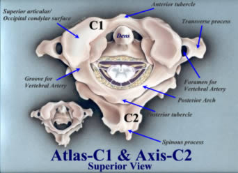
To understand what the 'anatomy of an atlas subluxation' looks like it's
first important to understand the anatomy of the atlas. The following
graphic depicts the atlas (C1) vertebra articulating with the axis (C2)
vertebra below it. The spinal cord with exiting spinal nerves can be
seen tethered by dentate ligaments to the inner margins of the vertebral
foramen. The human skull via the occipital condyles at the base of the
skull sits on top of (or articulates with) the superior articular condylar
surface of the atlas. According to Gray's Anatomy
[1] "This articulation
is a double condyloid joint. Its ligaments are the Anterior Occipito-atlantal,
Posterior Occipito-atlantal, two Lateral Occipital-atlantal and two Capsular." There
is a passage on each side in the Posterior Occipito-atlantal ligament
to allow penetration of the vertebral arteries and 1st cervical nerves.
The vertebral arteries exit the foramen in the atlas transverse processes;
penetrate the ligament and then loop up into the brain. A number of strong
ligaments support the skull and its contents and attach it to the cervical
spine. Many other ligaments and muscles enable movement, bending, twisting
and head rotation. There are other key structures within the vicinity
of the atlas and supporting ligaments. These are 4 pairs of cranial nerves,
namely the vagus, spinal accessory, glossopharyngeal and hypoglossal.
The common carotid arteries, which run underneath the sternocleidomastoid
muscle and jugular veins, are also present in the tissues of the neck.
At the base of the neck near the junction of the shoulders there are
some deep muscles known as the scalenes. These are the posterior, middle
and anterior scalenes. The anterior scalenes are of particular interest
as they are in very close proximity to the brachial plexus of nerves
and the origin of the vertebral arteries where they leave the subclavian
arteries. Even though the atlas is at the top of the cervical spine and
the above-mentioned structures are near the base of the neck, a subluxation
of the atlas and skull can result in these scalenes compressing the brachial
plexus and/or the vertebral arteries. This is due to altered weight bearing
from a shift in the centre of gravity of the skull, which in causes the
muscles of the head and neck to strain in maintaining the head upright
on the cervical spine. Also the phrenic nerve, which runs between anterior
and middle scalenes on its path to the diaphragm, can be likewise affected
by muscle and ligament compression and/or traction.
 Figure
1: Upper Cervical Spine – No subluxation
Figure
1: Upper Cervical Spine – No subluxation
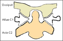
Figure1 opposite is a drawing of the upper cervical spine articulation
between the occiput (C0), the atlas (C1) and the axis (C2). The 'usual'
anatomy is for the occipital condyles (cream) of the skull to sit on
the atlas condylar surfaces and for the atlas lateral masses (white)
(shown cut away) to articulate with the facet joints of the axis (brown)
below it. The odontoid peg or dens of the axis should sit about central
to the foramen magnum. This is the front (anterior) view, if you were
looking directly at the face of this person. The double condyloid joints
are represented in this drawing by the 'gaps' between the occiput and
atlas lateral masses.
 Figure
2: Upper Cervical Spine – Skull to atlas subluxation
Figure
2: Upper Cervical Spine – Skull to atlas subluxation
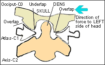
Figure 2 opposite shows what can happen to the 'usual' anatomy in Figure
1, when a force is applied to the skull on one side. The blue arrow indicates
the direction of trauma, which in this case is from the left side of
the person's head. This trauma can take many forms. In my case it was
my head striking the ground in a football tackle. For one little girl
it was falling out of bed and hitting her head on the bedside table and
for another lady playing basketball, it was a direct hit to the jaw.
The force is enough to push the skull sideways as it slides along the
atlas condylar surface. The occipital condyles of the skull can slide
off (over or underlap) the atlas condylar surface, however the anatomy
and the restraining ligaments limit this over and underlapping. The result
in this example is a head tilted to the left, and an atlas that's high
(elevated) on the right. The axis dens is no longer central to the foramen
magnum. See then animation
OCCIPUT
TO ALTAS SUBLUXATION to view the mechanism
of injury. The actual slope of the atlas condyles will determine whether
or not the atlas will actually rotate a little with respect to the occiput
(skull) and to the axis (C2). Flatter condyles will result in slight
rotation of the atlas, whereas steep angled condyles will most likely
not produce a great deal of rotation. The skull is quite heavy thus any
change in the centre of gravity will most likely produce undesirable
stresses on structures within the tissues of the cervical spine. This
situation has important implications for the spinal cord, which leaves
the foramen magnum and travels through the foramen (holes) in the atlas
and other skeletal vertebrae to the base of the spine. This also has
important implications for the vertebral arteries, which travel through
the cervical spine from C6 to the atlas, for the carotid arteries, which
travel beneath the sternocleidomastoid muscles, and for the cranial nerves
leaving foramen in the skull base. All of these structures have the potential
to be trapped, compressed, stretched or otherwise interfered with by
the taught muscles and ligaments involved in maintaining the skull and
its contents on top of the cervical spine. The resultant stresses on
arteries could yield reduction in oxygenated blood flow to the brain,
and on cranial nerves could result in attenuation of nervous system signals
to and from the brain. The cranial nerves which are potentially involved
are the vagus, spinal accessory, glossopharyngeal and hypoglossal. Of
course these arteries and nerves are of major importance to proper functioning
and maintenance of the human body.
 Figure
3: Even Death does not hide the proof!
Figure
3: Even Death does not hide the proof!
(Source: The Chiropractic Story silently told in a medical museum –
William G. Blair D.C., Ph. C., F.I.C.A., ©1973)
Copyright permission granted by Mrs Blair and Dr. E. Addington, Blair
Chiropractic Clinic, Lubbock, Texas, USA.
Website:
http://www.blairchiropractic.com
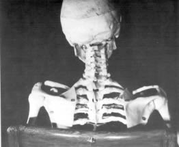
Figure 3 opposite is the skeleton of a soldier from the Spanish American
War (1898). He developed chronic rheumatoid arthritis, the disease progressing
and fusing most of his joints from his skull to his sacrum. His teeth
had to be removed in order to feed him since the rheumatoid arthritis
had also fused his jaw. Following his death and in accordance with his
will his body was given to the Army Medical Museum. His family honoured
his request and the skeleton is today displayed in the museum. Dr. William
G. Blair was given permission to examine and photograph the skeleton.
Figure 3, shows the head inclination as a result of a subluxation in
the upper cervical spine. Since rheumatoid arthritis does not progress
in death it is demonstrably obvious that the "spinal joints are
fixed in the same position they occupied at the time of fusion occurred" and "since
fusion can take place only in a living person, the present position of
these spinal vertebra in death are the same as they were during life".
These images are powerful and help to prove the existence of such subluxations
of the upper cervical spine, which are the focus of specific upper cervical
chiropractors like Dr. Blair and of my website. I suggest that it would
be interesting indeed to hypothesise that rheumatoid arthritis is one
sequelae of subluxations like this of the upper cervical spine. Fusing
of joints may well be the body's way of protecting the spinal cord, brainstem
and surrounding neurological and vascular structures from damage, by
allowing no additional movement, which would compromise them further.
Certainly it would not be the only disease process, which would be initiated
if someone sustained such a condition! This injury is so high up and
so close to the human central processing unit and other structures, as
mentioned earlier, that it's not hard to visualise such subluxations
may well be a root cause of human illness.
 Figure
4: A close look at the subluxation!
Figure
4: A close look at the subluxation!
Page 9 of Dr. Blair’s document
(The Chiropractic Story silently told in a medical museum – William
G. Blair D.C., Ph. C., F.I.C.A., ©1973) shows the photograph in Figure
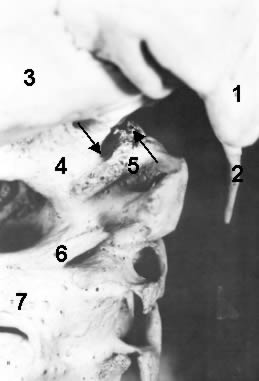
4 opposite. Let’s have a closer look at the anatomy of this subluxation.
The numbered structures are: 1. Right mastoid process; 2. Right styloid
process; 3. Right side
occipital
bone; 4. Right occipital condyle; 5. right lateral mass of the atlas
(C1); 6. Right side posterior arch of atlas; 7. Right lamina of axis
(C2).
According to Blair, "You will note the fusion of all three cervical
vertebrae. These vertebra are fused not only at the usual joints but
also at an unusual union of the posterior arch of first cervical (6)
with the right lamina of the second cervical (7). Since the growing together
process can take place only in the living body, every structure is fused
in the exact position it occupied during life.
Continuing our examination of Figure 4, note the two arrows. The arrow
on the left points to the outside (distal) margin of the right occipital
condyle (4). The arrow on the right points to the outside (distal) margin
of the right lateral mass of the first cervical vertebra (5). These are
the margins of the adjoining joint surfaces. Without misalignment the
two arrows would line up, instead there is displacement. The first cervical
vertebra is decidedly misaligned with the skull. By actual measurement
on the skeleton this displacement amounts to 3/8 of an inch (approx.
1cm). The second and third cervical vertebrae have followed the first
cervical vertebra to the misaligned position."
 Figure
5: An even closer look at the subluxation!
Figure
5: An even closer look at the subluxation!
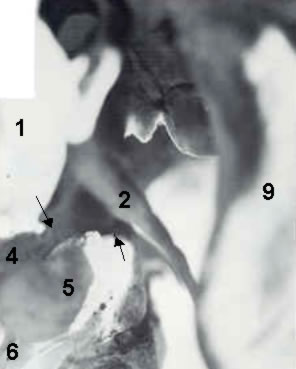
Page 10 of Dr. Blair's document shows these numbered structures: 1. Right
mastoid process; 2. Right styloid process; 4. Right occipital condyle;
5. right lateral mass of the atlas (C1); 6. Right side posterior arch
of atlas; 9. Right jaw (ramus of mandible). According to Dr. Blair, "Note
the two arrows. The arrow on the left points to the front (anterior)
margin of the right occipital condyle. The arrow on the right points
to the front (anterior) margin of the right lateral mass of the first
cervical vertebra. Again these margins are of adjoining joint surfaces.
If there were no misalignment, the two arrows would line up. Again there
is displacement. Again the first cervical vertebra is decidedly misaligned
with the skull. The actual amount of misalignment on the skeleton is
5/8 inch (approx. 1.6cm). Also note the bony fusion of first cervical
lateral mass, number 5, to the body of the second cervical vertebra below
and to the occipital condyle above, number 4. These structures having
grown together during life are found in exactly the same position after
death as they were during life. The body of the second cervical vertebra
has followed the atlas to the misaligned position and is aligned (juxtaposition)
with the atlas."
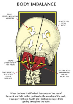
Figure 6 opposite provides information about
the effect an atlas subluxation will have on the surrounding ligaments
and muscles. In this case the
head is tilted to the right, the chin will be rotated to the left and
the neck will be in a left inflexion. The ligaments and muscles in the
sub-occipital area and those holding the head on to the cervical spine
will now be under stress and/or go into spasm. The spinal cord (brainstem
just above C1) will be 'kinked' at the craniocervical junction (skull
to atlas articulation). See the animation
KINK
SUBLUXATION to view the
mechanism of injury. The average human skull and its contents weigh in
at around 4.5 to 6 kg (approx. 10 to 14 pounds), and under normal circumstances
one can bend and rotate their neck without compromising the critical
neurological structures at this level. However, when this 4.5kg (10 pounds)
'bowling ball' is shifted the whole centre of gravity throughout the
cervical spine changes and this altered weight bearing will have negative
effects on neurological and vascular structures at the base of the skull
and throughout the neck, especially those structures which are in close
proximity to the muscles under stress. Remember that the vertebral arteries
and 1st cervical nerves pierce the posterior occipito-atlantal ligament
and it is highly possible that this and other ligaments will irritate
both these structures. The sub-occipital and cervical ligaments and muscles
implicated in this situation include but are not limited to the rectus
capitus posterior minor and major, obliquus capitis superior, obliquus
capitis inferior, sternocleidomastoid, leveator scapula, trapezius and
scalenes (in particular the anterior scalenes). Go to a bowling alley
and pick up a 10-pound bowling ball. Balance the ball on your fingers
and then tilt your hand so that the centre of gravity is now altered.
You will soon find out how hard it is to maintain this position for very
long. I know that in my own circumstance, my trapezius muscles were so
painful that I found it relieving to allow my head posture to go forward,
although over time the only relief was through putting my head down on
a surface or laying down in a bed. The stressed muscles and those in
spasm will begin to waste and over time this is very noticeable in sick
people. Their posture can be used as window into their health. There
is so much atrophy in the neck and shoulder muscles of people with obvious
atlas subluxations that part of the healing process or approach, after
the correction of the atlas subluxation, must include rehabilitation
of these muscles. This atrophy is very noticeable in people suffering
from Parkinson's disease, Multiple Sclerosis and other neurological diseases.
For further information on a suggested rehabilitation process for sufferers
of an atlas subluxation see my section
"A
NEW APPROACH TO WELLNESS"
 Figure
7: The Effects of the Atlas Subluxation
Figure
7: The Effects of the Atlas Subluxation
(Figure 7 reprinted with permission from Daniel
O. Clark D.C., Website:
www.uppercervicalillustrations.com).
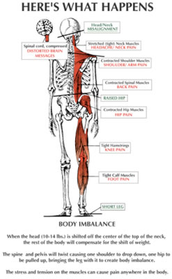
Figure 7 opposite explains what happens to your body if you unfortunate
enough to sustain a head injury which shifts your skull on your atlas
and results in a skull to atlas subluxation. Make no mistake for this
is exactly what happens. The ramifications of the subluxation can extend
to all parts of your body. In the most benign of these subluxations you
may only experience a little lower back pain and in the more serious
your whole body can be racked with pain and dysfunction. The tight sub-occipital
ligaments and cervical spine muscles can be responsible for headaches
or migraines as well as neck pain.
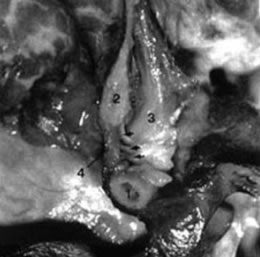 Figure 8: Myodural Bridge
Some years back Gary Hack D.D.S. et al 1995. "Anatomic relation
between the rectus capitus posterior minor and the dura mater" Spine
20 (23): 2484-2486 discovers a previously unknown ligament directly attaching
the posterior arch of the atlas to the dura mater of the brainstem and
cerebellum. This attachment has become known as the myodural bridge and
it is shown following in Figure 8.
The structures numbered in this dissection are as follows;
1. Posterior arch of the atlas; 2. Posterior occipital bone; 3. Rectus
capitus posterior
minor (RCPMI); 4; Dura mater of brainstem and cerebellum. The arrows
indicate the myodural bridge attaching the posterior arch of the atlas
to the dura mater of the brainstem & cerebellum. It is not difficult
to see that any misalignment of the atlas with respect to the skull could
traction the dura mater of the brainstem and cerebellum area. What would
be the effects of such traction? Certainly it is possible for headaches
and migraines to be an effect, but also there may well be other consequences.
Researchers at Michigan State University; "College of Osteopathic
Medicine" suggest "that
the function of the RCPMI muscle is to provide static and dynamic proprioceptive
feedback
to the CNS (central nervous system), monitoring movement of the head and influencing
movement of the surrounding musculature." In fact amongst other joints,
the joints of the upper cervical spine are rich in proprioceptors, which provide
positional feedback to the brain, in order for the brain to understand where
the joints and therefore the head are in space and to make appropriate adjustments
during movement. Close your eyes and point your first fingers on both hands.
Now try and touch the tips of these fingers without opening your eyes. Your
ability to touch your fingertips is an indication of your proprio receptive
abilities.
Any problems in this area may well be responsible for over and under corrections
made by your brain, which would manifest as dizziness or balance disorders.
Atlas subluxations, especially because they go hand in hand with some atrophy
of these
sub-occipital cervical muscles, could manifest as dizziness and/or balance
disorders. Meniere's Disease has been associated with cervical dysfunction
in a number of
medical papers, and I have personally witnessed and know of other people diagnosed
with Meniere's Disease who have had dramatic reversal of the disease's symptoms
following upper cervical correction to their atlas vertebra by specific upper
cervical chiropractors. This deserves further investigation as Meniere's Disease
and other balance disorders are highly distressing to sufferers.
Now looking at Figure 7 further you will note that because the skull
is tilted the pelvis will pull up on one side to compensate for the body
imbalance and pull that leg off the ground. This is known as a functional
short leg as opposed to an anatomical short leg. Of course, the leg is
not off the ground because the weight of the body keeps it on the ground
during walking. With the pelvis pulling up on one side the spinal column
will now twist into scoliosis. Now the back muscles are contracting on
one side which creates trigger points mainly at the 's' bend points in
the scoliosis, lower back pain is evident and now hip problems can be
a result. Leave this for long enough and the result may be surgical replacement
of a degenerated hip. Knee, ankle, groin and calf problems can be a direct
result, which people normally pass off as an aging thing or an accident.
For me this explains the back pain all the way down my back and my ankle
and knee problems, which corrected following correction of my atlas subluxation.
The brain is the key initiator of this lifting of the pelvis, in order
to align the pelvis and skeleton underneath the skull. There are cases
of scoliosis that have resolved completely or at least improved considerably
following upper cervical specific chiropractic correction of an atlas
subluxation. Re-position the skull on top of the atlas, and thus cervical
spine and the pelvis will be realigned directly underneath the skull.
Figures 9 & 10: NUCCA Types
Figures 9 & 10 reprinted with permission of National Upper Cervical
Chiropractic Association - see Website
www.nucca.org with special thanks
to Robert Goodman, D.C. NUCCA
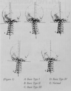
Figure 9 |
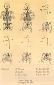
Figure 10 |
These graphics from the NUCCA site also show further the types of subluxations
that can occur at the upper cervical spine level.
Which one actually occurs in each individual really depends upon their
prevailing anatomy at that level and the direction and amplitude of the
force imparted during the trauma they received. While it is not always
the case, I have noticed that some people who have particularly large
misalignments in their upper cervical spine sometimes can exhibit only
minor symptoms, whilst others who have less of a misalignment can be
in a very bad state in terms of the symptoms they are experiencing.
UPPER CERVICAL ANIMATIONS
The Sid E. William Research Center
at Life University
www.life-research.edu [Note: this website is currently
unavailable] has some absolutely fantastic cervical spine animations
showing different motions and subluxations within the cervical spine.
I have chosen a couple of the animations to help explain further the
anatomy of the atlas subluxation and what happens to the occipito-atlantal
joint when there is a traumatic event. These animations follow herein.
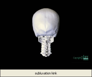 ANIMATION 1: The Kink Subluxation
Animation 1: Copyright permission granted by Life University with special
thanks to J. Roger Hinson, D.C.
This is known as the Grostic Kink Subluxation. This animation shows the
movement of the skull and atlas with respect to the lower cervical spine.

To play the animation RIGHT CLICK the PICTURE and click Linked Video
Clip Object and click Play Link or click on the following Hyperlink Kink
Subluxation.
Within the Media Player you can use the slider to move the animation
slowly through the subluxation. When you do this and leave the slider
in a position about halfway along you will now note the position that
the skull will be left in following a traumatic force to the skull, which
stretches the ligaments strapping the skull to the upper cervical spine.
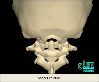 ANIMATION 2:
The Occipito-Atlantal
ANIMATION 2:
The Occipito-Atlantal
Animation 2: Copyright permission
granted by Life University with special thanks to J. Roger Hinson,
D.C.
This animation shows the movement of the skull with respect to the atlas
in lateral flexion to opposite sides. The skull slides with respect to
the lower cervical spine, and because of the skull weight and ligaments
its movement will drive and jam the atlas.
To play the animation RIGHT CLICK the PICTURE and click Linked Video
Clip Object and click Play Link or click on the following Hyperlink Occiput
to Atlas Subluxation.
Within the Media Player you can use the slider to move the animation
slowly through the subluxation. When you do this and leave the slider
in a position about halfway along you will now note the position that
the skull will be left in following a traumatic force to the skull, which
stretches the ligaments strapping the skull to the upper cervical spine.
Go to the Life University Research site and run the animations. You
will have a better idea, at least visually of what occurs.
www.life-research.edu [Note: this website is currently unavailable]
1. Gray, Henry (1977)
Gray’s Anatomy

 Figure
6: The Effects of the Atlas Subluxation
Figure
6: The Effects of the Atlas Subluxation
 The picture in the upper left corner above is a sagittal (side
view) of the brain stem with a cross section removed and layed out in
the main picture. In this picture the substantia nigra (long grey area
on either side of the cross section) is actually part of a system of
interconnecting nerve centers, called the
The picture in the upper left corner above is a sagittal (side
view) of the brain stem with a cross section removed and layed out in
the main picture. In this picture the substantia nigra (long grey area
on either side of the cross section) is actually part of a system of
interconnecting nerve centers, called the 


