Scarlet fever
From Wikipedia, the free encyclopedia
This article is about the disease. For Cee-Lo Green's backing band, see Scarlet Fever (band). For the song by Kenny Rogers, see Scarlet Fever (song).
| Scarlet fever | |
|---|---|
| Synonyms | scarlatina,[1] scarletina[2] |
 | |
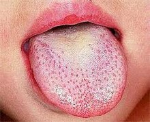
| Strawberry tongue seen in scarlet fever | |
| Classification and external resources | |
| Specialty | Infectious disease |
| ICD-10 | A38 |
| ICD-9-CM | 034.1 |
| OMIM | 012541 |
| DiseasesDB | 29032 |
| Patient UK | Scarlet fever |
Scarlet fever affects a small number of people who have either strep throat or streptococcal skin infections. The bacteria are usually spread by people coughing or sneezing. It can also be spread when a person touches an object that has the bacteria on it and then touches their mouth or nose.[1] The characteristic rash is due to the erythrogenic toxin, a substance produced by some types of the bacterium.[1][3] The diagnosis is typically confirmed by culturing the throat.[1]
There is no vaccine. Prevention is by frequent handwashing, not sharing personal items, and staying away from other people when sick. The disease is treatable with antibiotics which prevents most complications.[1] Outcomes with scarlet fever are typically good.[4] Long-term complications as a result of scarlet fever include: kidney disease, rheumatic heart disease, and arthritis.[1] It was a leading cause of death in children in the early 20th century.[5][6]
Contents
[hide]Signs and symptoms[edit]
Scarlet fever is characterized by:- Sore throat
- Fever
- Bright red tongue with a "strawberry" appearance
- Forchheimer spots (fleeting small, red spots on the soft palate)
- Paranoia
- Hallucinations
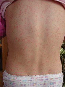
- A characteristic rash, which:
- is fine, red, and rough-textured,
- blanches upon pressure,
- appears 12–72 hours after the fever starts,
- generally begins on the chest and armpits and behind the ears but may also appear in the groin,
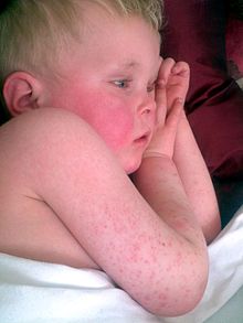
- on the face, often shows as red cheeks with a characteristic pale area around the mouth (circumoral pallor),
- is worse in the skin folds (so-called Pastia's lines, where the rash runs together in the armpits and groin, appears and can persist after the rash is gone),
- may spread to cover the uvula
- begins to fade three to four days after onset, upon which desquamation (peeling) begins. "This phase begins with flakes peeling from the face. Peeling from the palms and around the fingers occurs about a week later." Peeling also occurs in the axilla, the groin, and the tips of fingers and toes[7][not in citation given]
Rash[edit]
The rash is the most striking sign of scarlet fever. It usually appears first on the neck and face (often leaving a clear, unaffected area around the mouth). It looks like a bad sunburn with tiny bumps, and it may itch. It then spreads to the chest and back and finally to the rest of the body. In the body creases, especially around the underarms and elbows, the rash forms the classic red streaks known as Pastia's lines. On very dark skin, the streaks may appear darker than the rest of the skin. Areas of rash usually turn white (or paler brown, with dark complexioned skin) when pressed on. By the sixth day of the infection, the rash usually fades, but the affected skin may begin to peel.Other features[edit]
Usually, other symptoms help to confirm a diagnosis of scarlet fever, including a reddened and sore throat, a fever at or above 38 °C (100.4 °F), and swollen glands in the neck. Scarlet fever can also occur with a low fever. The tonsils and back of the throat may have a whitish coating, or appear red, swollen, and dotted with whitish or yellowish specks of pus. Early in the infection, the tongue may have a whitish or yellowish coating. Also, an infected person may have chills, body aches, nausea, vomiting, and loss of appetite.In rare cases, scarlet fever may develop from a streptococcal skin infection like impetigo. In these cases, the person may not develop soreness of the throat.
Course[edit]
When scarlet fever occurs because of a throat infection, the fever typically subsides within 3 to 5 days, and the sore throat passes soon afterward. The scarlet-fever rash usually fades on the sixth day after sore-throat symptoms started, and begins to peel (as described above). The infection itself is usually cured with a 10-day course of antibiotics, but it may take a few weeks for tonsils and swollen glands to return to normal.Pathophysiology[edit]
Scarlet fever is usually spread by the aerosol route (inhalation), but may also be spread by skin contact or by fomites. Although it is not normally considered a food-borne illness, an outbreak of scarlet fever due to infected chicken meat has been reported in China.[8]Asymptomatic carriage may occur in 15–20% of school-age children.[citation needed]
The incubation period is 1–4 days.
Microbiology[edit]
The disease is caused by secretion of pyrogenic exotoxins by the infecting Streptococcus bacteria.[9][10] Streptococcal pyrogenic exotoxin A (speA) is probably the best studied of these toxins. It is carried by the bacteriophage T12 which integrates into the streptococcal genome from where the toxin is transcribed. The phage itself integrates into a serine tRNA gene on the chromosome.[11]The T12 virus itself has not been placed into a taxon by the International Committee on Taxonomy of Viruses. It has a double-stranded DNA genome and on morphological grounds appears to be a member of the Siphoviridae.
The speA gene was cloned and sequenced in 1986.[12] It is 753 base pairs in length and encodes a 29.244 kiloDalton (kDa) protein. The protein contains a putative 30- amino-acid signal peptide; removal of the signal sequence gives a predicted molecular weight of 25.787 kDa for the secreted protein. Both a promoter and a ribosome binding site (Shine-Dalgarno sequence) are present upstream of the gene. A transcriptional terminator is located 69 bases downstream from the translational termination codon. The carboxy terminal portion of the protein exhibits extensive homology with the carboxy terminus of Staphylococcus aureus enterotoxins B and C1.
Streptococcal phages other than T12 may also carry the speA gene.[13]
Diagnosis[edit]

Streptococcus pyogenes (pictured)
Differential diagnosis[edit]
Cases need to be differentiated from Far East scarlet-like fever, an infectious disease first reported in the 1950s from Russia. Because of its similar clinical presentation to scarlet fever, it was initially thought to be caused by a Streptococcus. It is now known to be caused by a Gram negative bacillus—Yersinia pseudotuberculosis.Kawasaki's disease is another important differential, especially in its incomplete form. Scarlet fever appears similar to Kawasaki's disease in some aspects, but lacks the eye signs or the swollen, red fingers and toes. However, the signs of Kawasaki's disease may manifest over a few days, rather than at initial presentation. Complications of missed Kawasaki's disease are significant but rare, and include a 1–2% death rate and coronary artery aneurysms.
Prevention[edit]
Vaccines[edit]
No vaccines protect against S. pyogenes infection. A vaccine developed by George and Gladys Dick in 1924 was discontinued due to poor efficacy and the introduction of antibiotics. Difficulties in vaccine development include the considerable strain variety of S. pyogenes present in the environment and the amount of time and number of people needed for appropriate trials for safety and efficacy of any potential vaccine.[14]There used to be a diphtheria scarlet fever vaccine.[15] It; however, was found not to be effective.[16] This product was discontinued by the end of World War II.
Treatment[edit]
The treatment and course of scarlet fever is the same as that of strep throat.Antibiotic resistance[edit]
A drug-resistant strain of scarlet fever, resistant to macrolide antibiotics such as erythromycin, but retaining drug-sensitivity to beta-lactam antibiotics such as penicillin, emerged in Hong Kong in 2011, accounting for at least two deaths in that city—the first such in over a decade.[17] About 60% of circulating strains of the group A Streptococcus that cause scarlet fever in Hong Kong are resistant to macrolide antibiotics, says Professor Kwok-yung Yuen, head of Hong Kong University's microbiology department. Previously, observed resistance rates had been 10–30%; the increase is likely the result of overuse of macrolide antibiotics in recent years.Complications[edit]
The complications of scarlet fever include septic complications due to spread of streptococci in blood, and immune-mediated complications due to an aberrant immune response. Septic complications—today rare—include ear and sinus infection, streptococcal pneumonia, empyema thoracis, meningitis, and full-blown sepsis, upon which the condition may be called malignant scarlet fever.Immune complications include acute glomerulonephritis, rheumatic fever, and erythema nodosum. The secondary scarlatinous disease, or secondary malignant syndrome of scarlet fever, includes renewed fever, renewed angina, septic ear, nose, and throat complications, and kidney infection or rheumatic fever, and is seen around the 18th day of untreated scarlet fever.
An association between scarlet fever and hepatitis has been recognized for several decades.[18] The causal mechanism is unknown.
Epidemiology[edit]
This disease is most common in children ages 5–15; males and females are equally affected.[19][20] By the age of 10 years, 80% of children have acquired protective antibodies against streptococcal pyrogenic exotoxins, preventing development of scarlet fever.[20][21]History[edit]

Otto Kalischer wrote a doctoral thesis on scarlet fever in 1891.
The first description of the disease in the medical literature appeared in the 1553 book De Tumoribus praeter Naturam by the Sicilian anatomist and physician Giovanni Filippo Ingrassia, where he referred to it as rossalia or rosania. It was redescribed by Johann Weyer during an epidemic in lower Germany between 1564 and 1565; he referred to it as scalatina anginosa. The first unequivocal description of scarlet fever appeared in a book by Joannes Coyttarus of Poitiers, De febre purpura epidemiale et contagiosa libri duo, which was published in 1578 in Paris. Daniel Sennert of Wittenberg described the classical 'scarlatinal desquamation' in 1572 and was also the first to describe the early arthritis, scarlatinal dropsy, and ascites associated with the disease.
In 1827, Bright was the first to recognize the involvement of the renal system in scarlet fever.
The association between streptococci and disease was first described in 1874 by Billroth, discussing patients with wound infections. Billroth also coined the genus name Streptococcus. The organism was first cultured in 1883 by the German surgeon Friedrich Fehleisen. He cultured it from perierysipelas lesions. Rosenbach gave the organism its current name (Streptococcus pyogenes) in 1884.
Also in 1884, the German physician Friedrich Loeffler was the first to show the presence of streptococci in the throats of patients with scarlet fever. Because not all patients with pharyngeal streptococci developed scarlet fever, these findings remained controversial for some time. The association between streptococci and scarlet fever was confirmed by Alphonse Dochez and George and Gladys Dick in the early 1900s.
Nil Filatow (in 1895) and Clement Dukes (in 1894) described an exantematous disease which they thought was a form of rubella, but in 1900, Dukes described it as a separate illness which came to be known as Dukes' disease,[23] Filatov’s disease, or fourth disease. However, in 1979, Keith Powell identified it as in fact the same illness as the form of scarlet fever that is caused by staphylococcal exotoxin and is known as staphylococcal scalded skin syndrome.[24][25][26][27]
Scarlet fever serum from horses was used in the treatment of children beginning in 1900 and reduced mortality rates significantly.
In 1906, the Austrian pediatrician Clemens von Pirquet postulated that disease-causing immune complexes were responsible for the nephritis that followed scarlet fever.[28]
Bacteriophages were discovered in 1915 by Frederick Twort. His work was overlooked and bacteriophages were later rediscovered by Felix d'Herelle in 1917. The specific association of scarlet fever with the group A streptococci had to await the development of Lancefield's streptococcal grouping scheme in the 1920s. George and Gladys Dick showed that cell-free filtrates could induce the erythematous reaction characteristic of scarlet fever, proving that this reaction was due to a toxin. Karelitz and Stempien discovered that extracts from human serum globulin and placental globulin can be used as lightening agents for scarlet fever and this was used later as the basis for the Dick test. The association of scarlet fever and bacteriophages was described in 1926 by Cantucuzene and Boncieu.[29]
The discovery of penicillin and its subsequent widespread use has significantly reduced the mortality of this once feared disease.
The first toxin that causes this disease was cloned and sequenced in 1986 by Weeks and Ferretti.[12]
Dick test and vaccine[edit]

Gladys Henry Dick (pictured) and George Frederick Dick developed a vaccine for scarlet fever in 1924 that was later eclipsed by penicillin in the 1940s.
Gladys Henry Dick and George Frederick Dick developed a vaccine in 1924 that was later eclipsed by penicillin in the 1940s. Broth filtrates were used as the basis for the patent the Dicks took out on their vaccine in 1924 in the United Kingdom and in 1925 in the United States.[citation needed]
References[edit]
- ^ Jump up to: a b c d e f g h "Scarlet Fever: A Group A Streptococcal Infection". Center for Disease Control and Prevention. January 19, 2016. Retrieved 12 March 2016.
- Jump up ^ Shorter Oxford English dictionary. United Kingdom: Oxford University Press. 2007. p. 3804. ISBN 0199206872.
- Jump up ^ Ralph, AP; Carapetis, JR (2013). "Group a streptococcal diseases and their global burden.". Current topics in microbiology and immunology. 368: 1–27. doi:10.1007/82_2012_280. PMID 23242849.
- Jump up ^ Quinn, RW (1989). "Comprehensive review of morbidity and mortality trends for rheumatic fever, streptococcal disease, and scarlet fever: the decline of rheumatic fever.". Reviews of infectious diseases. 11 (6): 928–53. doi:10.1093/clinids/11.6.928. PMID 2690288.
- Jump up ^ Smallman-Raynor, Matthew (2012). Atlas of epidemic Britain : a twentieth century picture. Oxford: Oxford University Press. p. 48. ISBN 9780199572922.
- Jump up ^ Smallman-Raynor, Andrew Cliff, Peter Haggett, Matthew (2004). World Atlas of Epidemic Diseases. London: Hodder Education. p. 76. ISBN 9781444114195.
- Jump up ^ Sotoodian, Bahman; Rao, Jaggi (9 November 2015). "Scarlet Fever". MedScape. Retrieved 9 January 2016.
- Jump up ^ Yang, S. G.; Dong, H. J.; Li, F. R.; Xie, S. Y.; Cao, H. C.; Xia, S. C.; Yu, Z.; Li, L. J. (2007). "Report and analysis of a scarlet fever outbreak among adults through food-borne transmission in China". Journal of Infection. 55 (5): 419–424. doi:10.1016/j.jinf.2007.07.011. PMID 17719644.

- Jump up ^ Zabriskie, J. B. (1964). "The role of temperate bacteriophage in the production of erythrogenic toxin by Group A Streptococci". Journal of Experimental Medicine. 119 (5): 761–780. doi:10.1084/jem.119.5.761. PMC 2137738
 . PMID 14157029.
- Jump up ^ Krause, R. M. (2002). "A Half-century of Streptococcal Research: Then & Now". Indian J Med Res. 115: 215–241. PMID 12440194.
- Jump up ^ McShan, W. M.; Ferretti, J. J. (1997). "Genetic diversity in temperate bacteriophages of Streptococcus pyogenes: identification of a second attachment site for phages carrying the erythrogenic toxin A gene". J Bacteriol. 179 (20): 6509–6511. PMC 179571
 . PMID 9335304.
 - ^ Jump up to: a b Weeks, C. R.; Ferretti, J. J. (1986). "Nucleotide sequence of the type A streptococcal exotoxin (erythrogenic toxin) gene from Streptococcus pyogenes bacteriophage T12". Infect Immun. 52 (1): 144–150. PMC 262210
 . PMID 3514452.
 - Jump up ^ Yu, C. E.; Ferretti, J. J. (1991). "Molecular characterization of new group A streptococcal bacteriophages containing the gene for streptococcal erythrogenic toxin A (speA)". Mol Gen Genet. 231 (1): 161–168. doi:10.1007/BF00293833. PMID 1753942.
- Jump up ^ "Initiative for Vaccine Research (IVR)—Group A Streptococcus". World Health Organization. Retrieved 15 June 2012.
- Jump up ^ "Rudolf Franck - Moderne Therapie in Innerer Medizin und Allgemeinpraxis - Ein Handbuch der Medikamentösen, Physikalischen und Diätetischen Behandlungsweisen der Letzten Jahre". Springer Verlag. Retrieved 09 January 2017. Check date values in:
|access-date=(help) - Jump up ^ Ellis, Ronald W.; Brodeur, Bernard R. (2012). New Bacterial Vaccines. Springer Science & Business Media. p. 158. ISBN 9781461500537.
- Jump up ^ "Second HK child dies of mutated scarlet fever". Associated Press (online). 22 June 2011. Retrieved 23 June 2011.
- Jump up ^ Elishkewitz, K.; Shapiro, R.; Amir, J.; Nussinovitch, M. (2004). "Hepatitis in scarlet fever". Isr Med Assoc J. 6 (9): 569–570. PMID 15373323.
- Jump up ^ Czarkowski, M. P.; Kondej, B.; Staszewska, E. (2011). "Scarlet fever in Poland in 2009". Przegl Epidemiol. 65 (2): 209–212. PMID 21913461.
- ^ Jump up to: a b Zabawski Jr, Edward J. "Scarlet Fever: Epidemiology". Medscape. Retrieved 20 October 2014.
- Jump up ^ Czarkowski, M. P.; Kondej, B. (2010). "Scarlet fever in Poland in 2008". Przegl Epidemiol. 64 (2): 185–188. PMID 20731219.
- Jump up ^ Rolleston, J. D. (1928). "The History of Scarlet Fever". BMJ. 2 (3542): 926–929. doi:10.1136/bmj.2.3542.926. PMC 2456687
 . PMID 20774279.
 - Jump up ^ Dukes, Clement (30 June 1900). "On the confusion of two different diseases under the name of rubella (rose-rash).". The Lancet. 156 (4011): 89–95. doi:10.1016/S0140-6736(00)65681-7.
- Jump up ^ Weisse, Martin E (31 December 2000). "The fourth disease, 1900–2000". The Lancet. 357 (9252): 299–301. doi:10.1016/S0140-6736(00)03623-0. PMID 11214144.
- Jump up ^ Powell, KR (January 1979). "Filatow-Dukes' disease. Epidermolytic toxin-producing staphylococci as the etiologic agent of the fourth childhood exanthem.". American journal of diseases of children (1960). 133 (1): 88–91. doi:10.1001/archpedi.1979.02130010094020. PMID 367152.
- Jump up ^ Melish, ME; Glasgow, LA (June 1971). "Staphylococcal scalded skin syndrome: the expanded clinical syndrome.". The Journal of Pediatrics. 78 (6): 958–67. doi:10.1016/S0022-3476(71)80425-0. PMID 4252715.
- Jump up ^ Morens, David M; Katz, Alan R; Melish, Marian E (31 May 2001). "The fourth disease, 1900–1881, RIP". The Lancet. 357 (9273): 2059. doi:10.1016/S0140-6736(00)05151-5. PMID 11441870.
- Jump up ^ Huber, B. (2006). "100 years of allergy: Clemens von Pirquet—his idea of allergy and its immanent concept of disease". Wien. Klin. Wochenschr. 118 (19–20): 573–579. doi:10.1007/s00508-006-0701-3. PMID 17136331.
- Jump up ^ Cantacuzène, J.; Bonciu, O. (1926). "Modifications subies par des streptocoques d'origine non scarlatineuse au contact de produits scarlatineux filtrès". CR Acad Sci Paris (in French). 182: 1185–1187.
- Jump up ^ Dick, G. F.; Dick, G. H. (1924). "A skin test for susceptibility to scarlet fever". J Am Med Assoc. 82 (4): 265–266. doi:10.1001/jama.1924.02650300011003.
Further reading[edit]
- Rolleston JD (November 1928). "The history of scarlet fever". British Medical Journal. 2 (3542): 926–9. doi:10.1136/bmj.2.3542.926. PMC 2456687
 . PMID 20774279.
External links[edit]
| Wikimedia Commons has media related to Scarlet fever. |
- Scarlet Fever from PubMed Health


No comments:
Post a Comment