Saturday, 1 November 2014

Misalignment and Epilepsy

CESSATION OF A SEIZURE DISORDER: Correction of the Atlas Subluxation Complex

Robert J. Goodman, D.C., John S. Mosby Jr., D.C., M.D.

ABSTRACT

Observations of one patient presenting with a seizure disorder are reported. Relief of symptoms is

noted subsequent to correction of the misalignment of the occipito-atlanto-axial complex. The authors

suggest a relationship between the misaligned skull and subjacent vertebrae and some seizure disorders.

Key Words: epilepsy; atlanto-occipital joint

INTRODUCTION

The term epilepsy refers to any disorder characterized by recurrent seizures. Seizures are transient

disturbances of cerebral function due to abnormal paroxysmal neuronal discharges in the brain.

Approximately 0.5% of the U.S. population is affected. 1, 2


Epilepsy is grouped into two different etiological categories, idiopathic or constitutional and

symptomatic epilepsy. In idiopathic or constitutional epilepsy, seizures usually begin between 5 and 20

years of age. NO specific cause can be identified, and there are no other neurological abnormalities.1 The


causes of symptomatic epilepsy include abnormalities and perinatal injuries, disorders of metabolism,

trauma, space-occupying lesions, vascular problems, degenerative disorders, and infectious diseases. 1, 3-6


Clinically, seizures are categorized by description. The two major descriptive classifications are

partial seizures and generalized seizures. Partial seizures are determined by clinical observation and by

electroencephalograph manifestations. They affect only a restricted part of one cerebral hemisphere. In

simple partial seizures the patient remains conscious, but in complex partial seizures consciousness is lost.

Partial seizures may evolve into generalized seizures.1


Generalized seizures are categorized as petit mal or absence seizures, atypical seizures, myclonic

seizures, akinetic seizures, grand mal or tonic-clonic seizures, atonic or ionic seizures, and seizures that will

not fit into any other category.

Absence or petit mal seizures cause some reduction in postural tone, with some clonic or tonic

components. Consciousness is impaired. These attacks occur quickly. When the attack occurs as a person

is speaking, the person may miss a few words in mid-sentence then resume with the remainder of the

sentence when the attack subsides. If a child has a petit mal seizure while playing, for instance, he or she

may freeze, that is, stand perfectly still while reaching for a toy. These seizures will often cease at

approximately 20 years of age. Diagnosis is assisted by electroencephalographic studies which show

bilateral synchronous and symmetric 3Hz spike-and-wave activity.1


Atypical seizures are almost identical to the petit mal seizure except that changes in tone are more

dramatic and onset and termination of the attack is slower.1


Myclonic seizures cause single or multiple myclonic jerks and myofacial spikes are seen on an

EEG.1 Akinetic seizures present as a sudden loss of consciousness and EEG findings show synchronous

firing from deep lesions often in the frontal regions of the brain.7


The grand-mal or tonic-clonic seizure occurs with a sudden loss of consciousness, the patient

becomes rigid then falls to the ground and respiration is arrested for less than 60 seconds. This is described

as the tonic phase. The next phase is the clonic phase during which the body jerks violently for 2 to 3

minutes. Flaccid coma occurs next. During this type of seizure the tongue may be bitten and urinary or

fecal continence may be lost. The patient will then either recover consciousness, drift into sleep, or never

recover consciousness which is called status epilepticus.1


Atonic seizures are epileptic drop attacks where the patient, usually a child, loses all motor tone

and falls to the ground.8 The stigma of the helmet is often conferred of necessity on the patient with drop


attacks in order to protect him from further injury during these ictal events. These spells are often

intractable to treatment. Finally, the onset of ictal falling almost always occurs in patients who already

have one of more other types of seizures and implies a poor prognosis for the ultimate seizure control and

for normal mental development.9-11


Lennox-Gestaut Syndrome (L-G) is another classification of seizure disorder which has a peak age

of onset of four years.12 It is characterized by myoclonic and atypical absence seizures, regression of


intellectual functions and generalized spike wave discharges on the EEG at a rate below what is seen in

petit mal seizures. Several hundred attacks may occur in a day and multiple injuries from falls are

common. Diffuse cerebral atrophy is seen in 60% of cases.8


MEDICAL TREATMENT

The descriptive classifications above are important for determining the most appropriate medical

treatment. When patients have recurrent seizures, medication is prescribed until there have been no

seizures for at least four years. Epileptic patients are advised to avoid situations that could be dangerous or

life-threatening during seizures.1


All doctors must be aware of a condition called status epilepticus which is the rapid succession of

seizures so that the next seizure begins before the previous one has ended. Status epilepticus is a medical

emergency since continuous epileptic activity can damage the brain permanently. Of course death may

occur during a seizure if the patient aspirates contents of the stomach which either occlude airways or

prevent adequate oxygenation.8


When patients have been seizure free for at least four years withdrawal of medication may be

considered. There is unfortunately no way of predicting which patients can be managed without treatment.

Recurrence of attacks is most likely in patients who initially fail to respond to therapy, those with

convulsive jerking movements, those with multiple types, and those with continuing EEG abnormalities.1


HISTORY

Patient E is a five year old white female, who was small at birth and born breech. During her first

three years of life she had many viral infections and repeated attacks of otitis media. Some concern was

expressed by her mother over growth and language retardation. At age 13 months, however, psychomotor

skills had been assessed as normal.

At the age of 4 years 8 months, in October 1988, Patient E was playing as a day care center, when

she struck her head on the underside of a table. Within two hours her first grand mal seizure occurred.

Patient E’s second seizure occurred within three weeks.

Patient E was evaluated at the Mayo Clinic and was experiencing 10 to 30 seizures per day with

no seizure-free days. (Figure 1 is a graph prepared from Patients E’s daily seizure journal kept by her

parents. The recorded the number, time, type and severity of each seizure.) Seizure types were described as

being tonic, clonic, akinetic, and grand mal. The diagnosis of Lennox-Gestaut Syndrome was made. The

patient was described as being able to speak only a few intelligible phrases. She was tremulous, and had

difficulty standing.

Various laboratory tests were performed including a complete blood count, serum ammonia and an

SMA 20 and all were within normal ranges. A CT scan and MRI were negative for fractures and

pathologies. The EEG, however, showed a slow spike-wave abnormality as well as other features which

may occur in Lennox-Gestaut syndrome. The prognosis was determined to be grave and the comment was

made that the L-G syndrome is fraught with nearly uniform disappointment since medical therapy has very

few beneficial results.

Patient E was given Depakote, Zarontine, and ACTH therapy. Each was given singly and then

discontinued due to Patient E’s unusual or aggressive behavior. Finally, Tegretol/carbamazepine seemed to

have some positive effects on the grand mal type seizures. It was after this, however, that Patient E began

to have drop attacks.

On July 18, 1989 Patient E entered into a team evaluation and treatment plan at the Palmer

Chiropractic Clinic. At that time, Patient E was having 30 to 70 seizures per day. {Figure 1] Patient E

presented with a helmet, faceplate, and harness for protection against falls.

The patient’s communication skills were retarded. Physician exam revealed a waxy buildup in the

ears so that the TM could not be visualized. Neurological exam revealed the presence of hyper reflexive

and asymmetric reflexes. Orthopedic evaluation was unremarkable.

Further chiropractic evaluation revealed paravertebral muscular spasm in the cervical area;

cervical ROM severely restricted especially in the right lateral bending; right leg deficiency of 􀀁 to 1 inch;


suspected cervical misalignment resulting in an atlas subluxation complex.

A specific upper cervical x-ray series was taken.13,14 During patient placement for the nasium


view, certain postural deviations were noted. When asked to sit up straight, Patient E’s head, cervical, and

upper thoracic spine would not center directly over the pelvis and demonstrated excursion into the frontal

plane. No pathologies were noted on the x-rays.

Chiropractic x-ray analysis revealed a misalignment of the occipito-atlanto-axial region. Figure 2

is a representation of a radiograph showing Patient E’s misalignment in the frontal plane view. Rotation of

atlas in the transverse plane was also measured, but was minimal.

To correct the subluxation, Patient E was placed on the adjustment table side posture with the C1

transverse process as the contact point. An adjusting force was introduced to the spine using specific upper

cervical adjusting procedures.15 The success of the adjustment was measured by lessening of leg disparity,


increase of pelvic resistance, postural changes, and finally post adjustment x-ray analysis.

Patient E was adjusted on three consecutive days. After the first adjustment, Patient E’s right leg

changed from one inch deficiency to no noted deficiency.16 The patient began to rub her eyes and seemed


drowsy after the adjustment. The seizure pattern remained quite high that day. On the second day, the preadjustment

leg deficiency was 1/4th inch on the right. After the adjustment, the legs were even and the


pelvic resistance on the right was stronger that the day before. The adjustments were always made between

9:00 am and 1:00 pm. After 1:00 pm on the second day, Patient E had no more seizures during the day.

On the third and final day of care at the Palmer Chiropractic Clinic, the leg deficiency was 1/8th inch.


Again, after the adjustment, the legs were even, and the pelvic resistance was equal bilaterally. Patient E

had no further seizures after 1:00 pm on this day. The parents of Patient E commented that she was

showing more energy and more stability when standing. A postural change was quite evident when Patient

E was prepared for the post x-ray series. When instructed to sit up straight, Patient E’s head and upper

spine were centered over her pelvis. The vertical centering lines on the bucky were used for comparison.

Post x-ray analysis revealed a 91% reduction of the misalignment factors with all structures centered on the

vertical axis.

The parents had been counseled that after an adjustment they could expect exacerbations or

changes in symptomatology on the 3rd, 7th, 14th, and 28th day.17 On the 17th day after the adjustment

procedure, the seizures numbered almost 100 (more than ever before). On the 27th day, the seizures abated.


[Figure3] The seizures remained absent for approximately four weeks. The carbamazepine dosage was not

changed during this period.

At the time of this report, the dosage had been reduced by almost one half for the past two weeks.

Patient E has had six or fewer seizures per day. The seizures are completely absent on some days. After

fewer than 60 days of chiropractic care Patient E has been speaking with five or six word sentences. A

current speech evaluation is pending from her therapist and our patient has not been adjusted since July

1989. At re-evaluation, Patient E had no leg deficiency and her condition continues to improve.

DISCUSSION

The authors could find little chiropractic literature pertaining to epilepsy or seizure disorders.

Young reported on three cases which responded to chiropractic care.18 IN the medical articles reviewed on


this subject, little hope, if any, is expressed for the control or cessation of childhood seizures of this type.

Using statements like “woefully”, “poor diagnosis”, “retractability to treatment”, “fraught with uniform

disappointment”, and “grim foreboding”,2 this literature demonstrates the need for research no only in


alternative methods of treatment, but also research into possible causative factors as well.

The remission of Patient E’s epilepsy is a phenomenon whose timing is shortly preceded by the

adjustment procedure. Perhaps not all seizure disorders can be helped with spinal adjustments. However,

chiropractic care would surely be the most conservative treatment in the primary care system. Careful

investigation into each case may reveal possible avenues that may eventually help the patient reduce or in

some cases entirely eliminate the need for drug therapy.

ACKNOWLEDGEMENTS

The authors wish to acknowledge George Hess, D.C., Chris L. Hendricks, D.C., Vern Hagen,

D.C., and Dale Strama, D.C. for their part in the care of Patient E and Alana Ferguson, M.S., Susan Larkin,

D.C. and David Guerriero for help in manuscript preparation.

REFERENCES

1. Schroeder SA, Krupp MA, Tierney LM. Current medical diagnosis and treatment. Norwalk,

CT:Appleton & Lang; 1988: 576-580.

2. Snead OC III. Epilepsy in children, a practical approach. Seminars in Neurolgoy 1988; 8(1):24-

34.

3. Allen RB. Nutritional aspects of epilepsy. International Clinical Nutrition Review 1983; 3(3):3-

10.

4. Recent advances in childhood epilepsy. 17th International Epilepsyn Congress. Epilepsia 1988;


29 Supplements 3:S1-S24.

5. Sun JY. An analysis of the causes of epilepsy in 440 cases. Chung Hua Shen Ching Ching Shen

Ko Tsa Chih 1988 June; 21(3):141-4.

6. Crowell GF, Roach ES. Pyridoxine-dependent seizures. Am Fam Physician 1983; 27(3): 183-

187.

7. Harvey AM, Johns RJ, McKusick VA, Owens AH, Ross RS. Principles and practice of medicine.

Norwalk, CT; Appleton-Century-Croft; 1984:1236

8. Andreoli TC, Carpenter CCJ, Plum F, Smith LH Jr. Cecil essentials of medicine. 2d.ed.

Philadelphia: W.B. Saunders; 1990.

9. Evans OB, Hanson RR, Snead OC. The primary generalized epilepsies of childhood. Semin

Neurol 1988 Spring; 8(1):12-21.

10. Pazzaglia P et al. Drop attacks: an ominous change in the evolution of partial epilepsy.

Neurology 1985; 35: 1725-30.

11. Markland ON. Slow spike-wave activity in EEG and the associated clinical features; often called

Lennox-Gastaut Syndrome. Neurology (Minneap) 1977;27:746-757.

12. Laidlaw J, Richens A, Oxley J (eds). Textbook of epilepsy. 3d ed. New York; Churchill

Livingstone 1988.

13. Gregory RR. The NUCCA course: adjusting the atlas subluxation complex. Monroe, MI:

National Upper Cervical Association Inc. 1988.

14. Dickholtz M. Patient alignment for upper cervical x-rays. Upper Cervical Monograph 1980;

2(8):2-7.

15. Gregory RR. The NUCCA course: adjusting the atlas subluxation complex. Monroe, MI:

National Upper Cervical Association Inc. 1988.

16. Gregory RR. Biomechanics of C1 subluxation production. Upper Cervical Monograph 1988;

4(5).

17. Gregory RR. Post-adjustment healing cycle: nerve and tissue repair. NUCCA Educational

Conference, May 1979, unpublished seminar notes.

18. Young G. Chiropractic success in epileptic conditions. ACA Journal of Chiropractic 1982


MS lesions of the Cervical Cord - Upright health


MS Lesions of the Cervical Cord

Axial FLAIR (a, b & c) and T2 weighted (d) Bra...
Image via Wikipedia
                                                                                                     Multiple Sclerosis is technically a neurodegenerative disease of the brain. Classically MS is associated with two or more supratentorial (above the covering over the cerebellum) periventricular hyperintensity signals. The lesions are the white spots on the brain scan on the right. The large shadows in the middle of the brain shaped like a butterfly are the lateral ventricles. The periventricular area surrounds the ventricles. In addition to the brain, however, some MS patients also get lesions in the cervical cord.
Demyelinating type lesions in the cervical cord without lesions in the brain are not considered to be classic signs of MS. Lesions below the cervical cord are, likewise, not considered to be MS. Instead they are given different names.
In this post I will stick to MS lesions in the cervical cord and disregard lesions of the lower cord such as those seen in amyotrophic lateral sclerosis (ALS), primary lateral sclerosis, Devic’s Disease (neuromyelitis optica) and others. I will also ignore the lesions sometimes associated scoliosis and abnormal curvatures of the spine.
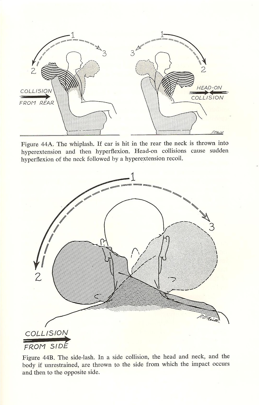
According to Schelling, the lesions seen in the cervical cord in MS are due to stretch and shear stress. Many types of trauma, such as whiplash in the picture on the left, cause hyperflexion and hyperextension of the spine. The strain can occur in a front to back motion or from side to side. At the same time, whiplash type traumas also strain the spinal cord inside the spinal canal. 
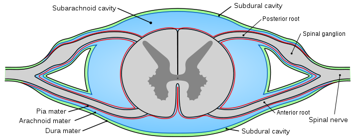
 The spinal cord, like the brain has three protective coats called meninges. The outer coat is called the dura mater. The middle coat is called the subarachnoid mater and the inner coat is called the pia mater. Except for the attachment of the tail end of the cord, called the filum terminale, to the tail bone, called the coccyx, the spinal cord moves freely, unattached inside the spinal canal.


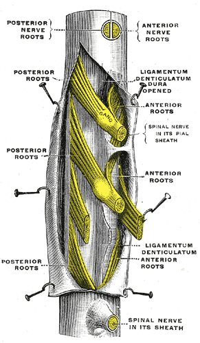
The three coats are held together by denticulate ligaments that attach the inner pia mater to the outer dura mater at the nerve roots.  Click on the image on the right for a closer look. The subarchnoid space lies between the subarchnoid mater and the pia mater and contains a significant volume of cerebrospinal fluid, called CSF. There is also lymph like fluid that serves as a lubricant between the outer coat of the dura mater and the middle coat of the subarachnoid mater. This allows the inner contents of the cord to move somewhat independently to a limited degree.
According to Schelling, mechanical strains of the spine can take the cord to extreme limits of stretch. It can also cause shear stress within the cord between the pia mater on the inside and the dura mater on the outside. The stress points occur at the attachment of the denticulate ligament. Furthermore, violent back jets of CSF in the subarachnoid space amplify the strain like a boat moored to a dock hit by hurricane force waves. The waves amplify the strain and rip the ropes and boats from their moorings. Similarly, violent waves of CSF flowing through the subarachnoid space amplify the strain acting on the denticulate ligaments. When they reach a critical threshold, engineer’s call the yeild point, the denticulate connective tissue attachments of the pia mater to the dura mater will tear. The strained tissues of the pia mater further damage the meylinated nerve tracts they cover. Click on the image above to get a closer look. This is a cross section of the cord and shows the inside. The H shape in the middle of the cord is the grey matter. The myelinated white fiber tracts surround it. The blue area is the subarachnoid space which contains CSF.

The lesions seen in the cervical cord are micro tears in the meninges and nearby myelinated nerve pathways . The tears cause scars  to form called sclerotic plaques.  Because they occur on the outside along the flanks of the cord, the tears tend to effect the outside structures of the cord.
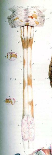
The outside of the cord (see picture to left)contains the long myelinated high speed primary nerve pathways of the central nervous system. Primary nerves go from the brain to particular destination levels in the cord. The long white tracts on the outside of the cord contain both sensory nerves for detecting sensations related to the environment, muscle tone and balance, and motor nerves for running the muscles. Tears in the area of the denticulate ligaments thus affect nearby myelinated nerves in the cord resulting in muscle weakness and warped sensations called paresthesias.
In brief, according to Schelling, one of the likely causes of the lesions of multiple sclerosis seen in the brain are due to violent venous back jets associated with trauma.  The lesions seen in the cervical cord are due to severe overstretching and shear stresses within the cord itself magnified by hydraulic stress from CSF waves. There is still more to the story, however, that needs further exploration and explanation.  That’s where my theory may help to fill in some of the missing pieces to the puzzle.
My theory is that craniocervical syndromes can cause chronic edema (swelling), ischemia (decreased blood flow) and NPH (normal pressure hydrocephalus) conditions in the brain. Similar events can occur in the cord due to abnormal conditions of the spine. These chronic conditions can, in turn initiate neurodegenerative processes such as the glutamate cascade, which follows strokes.
In my next post I will discuss the posterior blood supply routes to the brain and circulation to the cord. The veins are not the only problem in MS. The arteries are a big part of the problem as well. In addition to MS, I suspect that certain cases of sclerosis of the long myelin tracts of the lower cord, such as ALS and PLS are probably due more to chronic ischemia. This is probably why lesions sometimes also show up in cases of abnormal curvatures of the spine such as scoliosis and kyphosis. Oxidative stress and chronic ischemia are chief suspects in the cause of demyelination and the shape of the spine, and thus the spinal canal, affect arterial and venous blood flow in the cord.
For additional information on this and related topics visit my website at http://www.upright-health.com.

CCSVI and brain pressure - Dr Flanagan

CCSVI and Brain Pressure

CCSVI and CSF
Cranial hydrodynamics is a term used by radiologists to describe fluid mechanics in the brain, which includes blood and cerebrospinal fluid called CSF. Intracranial compliance is term used to describe pressure changes in the brain inside the skull caused by changes is CSF pressure as it pulsates on its course through the brain being driven by waves arising from the heart and arterial pulse waves along with respiratory waves, which amplify them. Techically they are called cardiorespiratory or B waves. Some scientists also call them volume waves in the brain.
In brief, the brain pulsates just like the beat of the heart. Things that disturb intracranial compliance and the rhyhmical movement of fluids can have profound affects on the health of the brain because it lies within a somewhat restricted closed container. The other two elements within the container are blood and cerebrospinal fluid mentioned above. An increase in the volume of any one element within the cranial vault must result in a decrease of one or both of the other elements. Two of the elements, blood and CSF are similar to water and therefore not very compressible. The brain, on the other hand is made of billions of nerves and over fifty percent fat, so it is much more compressible than blood or CSF.
CSF produced in the chambers of the brain called ventricles. A dense network of blood vessels surround the ventricles. CSF is made from blood that is drawn from the network of blood vessels through an extra-fine fine filter, called the blood brain barrier. The blood is drawn by a concentration created by salt, as well as a pressure gradient created by upright posture.
CSF is used to support the brain, which floats inside the skull as well as for cushioning and protection. Moreover, the brain has no lymph system. Instead, waste products are removed through the CSF system, which empties into the main vein at the top of the head called the superior sagittal sinus.
CSF production, flow and drainage from the brain is inexorably linked to venous drainage from the brain. CSF pressure in the ventricles is normally slightly higher than venous pressure so that it’s flow is toward the veins.
Upright posture creates a slightly negative pressure in the superior sagittal sinus, the main vein at the top of the head. This draws venous blood and CSF toward it. From here blood flows downhill along a steep slope like a waterfall to the base of the skull. In the bottom of the skull drain into either the jugular or vertebral vein routes.
The CSF pressure gradiant is very small so that venous drainage issues, such as back pressure in the veins in the basement of the skull, can affect intracranial compliance. It can also affect cranial hydrodynamics resulting in either too much CSF in the brain, called normal pressure hydrocephalus in adults or too little. Moreover, a decrease in the volume of CSF decreases brain support and raises the risk of pressure conus and Chiari like conditions. I cover cranial hydrodynamics thoroughly in my book, as well as Chiari and pressure conus conditions.

2 Responses to CCSVI and Brain Pressure

  1. narnia says:
    great post, just the kind of information I was looking for
  2. Amy Jewell says:
    I love your site! I am amazed. My daughter had viral meningitis and renal failure in 2005. Since then she has had ICP, many misdiagnosis’ leading to adrenal insufficiency. But ever symptom since then, migraines, fluid ears, chronic viral respiratory infections every winter never fit with adrenal problem. She has always been able to make cortisol-but never pass a stimulation test. All other hormones test fine. She is on physiological dose of 15 mg total day of cortef. Finally, 2008 she was diagnosed with POTS. The doctor added Nadolol to her meds, 3000mg salt, compression stockings. But starting last fall, migraines have grown worse, she has had esophageal candida 3 times and since december her head pain is stuck every day-all day. Pain gets worse as day goes on. I would love to get standing MRI for her. I get migraines, I can feel these lumps and bumps on my skull, but they go away. Kims skull feels like that all the time. How do I get her help? She gets new neuro doctor next week. What do I say without sounding like crazy mom with “internet” knowledge?

Leave a Reply


Whiplash injuries and CCSVI - Dr. Flanagan/Uprightdoctor

Whiplash Injuries and CCSVI

Upper Cervical Spine
In engineering terms, stress from any type of force always strains the structures and materials they act on. Any structure or material can be strained technically speaking. All strains by definition result in deformation of their respective structures and materials. The difference is that elastic strains return back to normal while plastic strains do not. In engineering the crossover point between elastic and plastic strains is the yield point. Strains are categorized as large and small strains, which can be further broken down into infintissimal and micro type strains. Regardless of size, even small strains can can have a large impact on sturctures like cracks in a foundation fortelling of catastrophic failure.
Jumping from a steel bridge while attached to a bungee cord causes large elastic strains and deformation in the bungee cord. At the same time it causes infintissimally small elastic strains in the bridge and small but larger, hopefully elastic strains in the jumper’s body. On the other hand, whiplash and other suddden jerky or even slow sustained types of injuries to the spine can cause chronic micro plastic types of strains.
Chiropractors collectively catergorize all mechanical strains of the spine as subluxations. Technically speaking, subluxations are micro mechanical plastic strains of the spine that cause dis-ease in physiology, which is function. Some chiropractors argue that subluxations must include interference to communication in the nervous system, and that the only place where interference can occur is in the upper cervical spine. It is an age old arguement in chiropractic.
All strains of the spine, however, alter physiology. Furthermore, the spine is a structure, not a bunch of isolated segments so that one faulty segment can impact the function of the whole structure. Strains of the upper spine can affect the lower spine just as strains of the lower spine can affect upper areas. A spine that lists due to scoliosis from leg length discrepancy affecting its base can affect the upper cervical spine. Likewise a ten pound head that lists to one side can affect segments much further below, including the low back and legs. Furthermore, abnormal curvatures of the spine have been shown to affect the contents of the spinal canal, especially the vertebal veins, as they get compressed up against the inside curve of the canal, which can result in venous congestion and sluggish blood flow in the area. Lastly, deformation of the cartilage of the spine, called discs, can compress nerve roots directly, while other types of spinal strains deform soft tissues tunnels such as thoracic outlets in the shoulder girdle and the femoral and sciatic foramen in the pelvis.
The upper cervical spine, however, is without question one of the most critical areas of the spine, subject to a great deal of wear and tear stress that starts with birth and sometimes results in chronic strains called subluxations. The differece between upper cervcial strains and those that occur further down in the spine is that upper cervical subluxations, that is strains, affect the health of the brain and cord.
Among other things, upper cervical subluxations are associated with deformation, albeit small, of hard and soft tissue tunnels that contain critical circulatory routes for blood and cerebrospinal fluid flow, including venous drainage routes used to drain the basement of the brain during upright posture. Thus, UC subluxations can result in CCSVI. But they also do much more. The fact of the matter is, UC subluxations don’t simply pinch nerves, as chiropractors like to say, nor do they simply interfere with communication between the brain and the body. In contrast to a fracture of the UC spine that can kill a person instantly, upper cervical subluxations slowly strangle the life out of the brain and cord.

18 Responses to Whiplash Injuries and CCSVI

  1. Jean says:
    I had my first MS attack in July, 1992. It was not formally diagnosed RRMS until September, 1993 by a neurologist. My second attack was in October, 2004. By 2001, my MS had graduated to SPMS, but I had reached a plateau until October, 2006 when I was rear-ended at 50 mph making a left hand turn by an uninsured driver. Since then, my MS symptoms went downhill. I’ve read that whiplash can cause soft tissue injuries, but I developed new pains including neck & shoulder pain, knee pain (both), low back and abdominal pain. My auto insurance co. denied medical claims due to a “pre-existing condition” -MS. Since the accident, I’ve also developed scoliosis per x-rays. MRI’s showed some degeneration of the spine. Any recourse or treatment? I’ve tried chiropractic, physical & occupational therapy, Osteopathic Manipulative Therapy, even acupuncture, but I’m still in daily pain. My attorney wants to settle through mediation to avoid a less educated jury. I just want the pain to stop. I have considered being tested for the CCSVI which is how I found your site. Any thoughts?
    • Drs Flanagan says:
      Hello Jean, It’s medicolegal injustice backed up by bogus epidemiological studies the AMA uses to discredit a connection between MS and trauma. The poorly designed studies provide defense attorneys false evidence to protect their clients and deprive victims of just compensation. The AMA continues to maintain it’s position, not to protect patients, but to prevent a possible connection to trauma that might compete with precious pharmaceutical products and corporate profits. The attorney’s typically hire hack neurologists with little or knowledge about the subject. I would love see them explain in court to a jury with a little common sense why overstretching the brain and cord and massive venous and CSF backflows in the brain and cord can’t be a cause MS.
      There is a limit to what any professional can do to repair badly damaged tissues. CCSVI testing and treatment is definitely a consideration if corrective care of the spine fails. That said, what type of chiropractic care did you get? Aside from musculoskeletal complaints do you have any neurological signs and symptoms?
  2. I found these items most interesting, I had a motoring accident in 2006, since that time I have been sent to see the so called Medico-legal experts. One informed me that I had had two replacement hips. “News to me”. another one of these highly mobile specialist
    medical records scrappers who travel the country trying to find a pre-existing condition to use in order to get your insurers out of paying compensation ; his examination lasted only 7 minuets, he told me on entering the room that he had almost completed the report
    without seeing me.
    I would like to find others who have had problems with Law firm and these low grade
    Medico-legal experts. Can you put me in contact?. supply my email address if you wish.
  3. I am a 41 year old female that has been disabled for more than 3 years. There has been difficulty is diagnosing and my symptoms continue to get worse with activity. I had bad whiplash in 1988, ‘recovered’ from that then started having issues with lifting, arm numbing, headaches, etc. Saw a chiropractor for a few years for my neck and mid-back which seemed to help at the time. Since then I have had whiplash two more times; in Aug. 2006 (mild) and Aug. 2007 (medium). In 2007 I had a water tubing accident where I fell from the tube at high speed and hit the water with my right side so hard that I felt a numb feeling from my waist down for about 1 minute. I seemed to recover from that but starting in summer 2008 I started having great difficulty with expanding my ribcage without severe pain, headaches and the list is long. It progressed over time and now I am left disabled without an answer. Most of my pain seems to be triggered by the use of my right hand. Grasping objects is the worst. Writing will set off muscle spasms all down my right side and I can barely walk after. Not much is showing up on various tests that I have had and I am very frustrated. Has anyone else had this effect from multipe whiplash? I have a lot of joint pain, headaches (back and left side of head), pins and needles in my arms if I sit in one place for 5 minutes of more or when sleeping on my side, various pressure points all over my ribcage and neck, weakness in my right hand and arm, difficulty breathing from rib pain, muscle spasms on right side of body, loss of temp sensitivity in hands, etc.
    I have been tested for MS and it came back negative but when they did the MRI for that I had been having a ‘better’ day than normal because I did not do activities before. I am desperate for some answers and would like to get my life back to normal. This has been devestating to not have answers.
    Thank-you
    Teresa
    • Hello Teresa,
      Your symptoms are due to pressure on the brainstem and cord most likely caused by upper cervical misalignment. It is impossible to imagine how you could not have injured your cervical spine, especially after the water tubing injury where you felt a “numb” feeling from the waist down for a minute. I don’t know what particular tests were ordered but I suspect that the tests were negative mostly because you don’t have any lesions on brain scans. That doesn’t mean you don’t have serious symptoms similar to MS and you are progressing. A recent study by Dr. Damadian the inventor of MRI showed that there is a connection between MS and trauma. More and more evidence is pointing that way. I would strongly recommned you consult with a highly qualified upper cervical chiropractor to take specific x-rays of your upper cervical spine and get it corrected.
    • Nanette says:
      Hello. I really thought that I was out of my mind, because I do not think people understand what pain can do to you. I had a rear end accident beginning of this year. Did not think anything about it, accept for being without my car for about 3 weeks. I had neck pain the evening, but nothing after that. Only about 3 to 4 weeks I started having servere head aches, neck pains – as soon as I sit for to long this will start. I have been to 4 different docters, neuroligst / surgeons, chiros, physio, you name it. I have lost my job because I can not sit for longer than 20 / 30 minutes before the pain get so servere that I cannot focus and my bp gets sky high. This is now going for the 7th month and I still have the same symptoms. Headaches, neck pains, shoulder. Left side goes numb. my face (only left) has got this tingling feelling, my toes go numb if I walk a long distance. I feel so frustrated because I cannot do normal house hold chores as well. I have to stand most of the time and this is leading to extreme lower back pain. I am starting to have memory loss, difficulty spelling and concentration is 0. I also started having problems with my bladder and my bowel pains. It is just getting worse. So I fully do understand how you feel. The worst is NOT getting any answers. All my test came back as normal = but believe me I am far from NORMAL!!
      Nanette
      • Hello Nanette,
        The rear end collision is the cause of your problems. You clearly had a whiplash injury that resulted in neck and shoulder pain with headaches. The problem started seven month ago and progressed to a chronic strain causing worsening of signs and symptoms. The strain on the muscles and connective tissues of the cervical spine are affecting the blood vessels and nerves in the base of the skull which is being transmitted to tissues inside the cranial vault. I don’t know what type of chiropractor you saw but you need a better evaluation of your spine, which you won’t get from a neurologist, orthopedic surgeon or a mediocre chiropractor. Something is clearly wrong and the doctors are obviously incapable of finding it due to incomplete or poor examinations. I highly doubt that you have no abnormal findings. Among other things, I have no doubt that you have inflammed sensitive soft tissues and probably faulty range of motion in your cervical spine. You may also have torn ligaments and connective tissues. Someone is not looking. So keep looking until you find someone who does. Now is the time to get it fixed before more damage is done.
  4. Mario says:
    Dr. Flanagan thanks for creating this site and for providing feedback. This is all very interesting to me as was diagnosed with RRMS in 2004 after sensory activity in face and primarily the left side. History: Head injury as child fell off a step and landed on hydro meter split chin open some stitches….11 yrs old car accident 27 stitches right top side of head…me on pedal bike…drunk in truck….knocked unconscious. 19 yrs old tunnel board (boogie board) behind ski boat hit water hard and major whiplash….chiropractor same day. Since then have had lots of neck fatigue, stiff necks and pain off and on over the years. Xray shows some compression and surgeon said not a candidate for surgery as was not bad enough then MS was diagnosed so the neck is no longer even looked at. 2 lesions on c-spine and they think one in brain but not 100% certain.
    Seems to me you are onto something…the issue for me as now that I have been diagnosed chances of getting anyone to open this up again is nil. What would be your approach and suggested treatment?
    Having an informed intelligent suggestion when asking seems to get more action so any suggestions on how to broach it would be appreciated.
    • Hi Mario,
      Your welcome. Basic cervical x-rays would be helpful to check for spondylosis and curvature problems. Specific upper cervical x-rays would also be helpful. In addition to x-rays, upright cervical and craniocervical junction MRI scans along with flexion and extension cervical views would be helpful. Once again, the scans should be done to check for spondylosis, abnormal curvatures and damaged connective tissues, espeically in the craniocervical junction. Phase contreast cine MRI would be further helpful to check blood and CSF flow. My suggested treatment would be based on what the x-rays and cervical scans show.
  5. Ragen says:
    Dr. Flanagan:
    Have you seen situations where trauma (in my case, being thrown from a horse) that resulted in concussion lead into orthostatic headache? It was suspected that I had a csf leak, but no enhancement has shown on MRI imaging and I had no relief from epidural blood patch.
    • Hello Ragen,
      The head is connected to the upper cervical spine. Except in rare cases, trauma to one typically affects the other. Since you showed no CSF leaks and no relief from an epidural blood patch, your orthostatic headache is obviously not due to CSF leaks causing intracranial orthostatic hypotension. Your headache is most likely due to muscle, joint and connective tissue injuries to the spine caused by the fall.
  6. L.E. says:
    I can feel a bone out of alignment in my upper neck right at the base of my skull. I had a severe whiplash injury about 20 years ago and another whiplash injury just 2 years ago. I have migraines, numbness/tingling down left arm and weak grip, limited range of motion in neck, tight muscles in neck and shoulders which feel like “steel” according to the Physical Therapists I have been to. I also suffer from a constant internal buzzing/vibration, dizziness/fuzzy-headed (and always left side), sinus issues, plus ongoing trouble with stomach which one dr suggested was triggered by vagus nerve, but didn’t offer any real solution to it. The xrays I had after most recent car accident didn’t show anything. I have been to a chiropractor who said he can feel the misalignment–but no lasting relief from treatment. Had an MRI that didn’t show anything significant. I have been told from a CT done for another problem that I do show degenerative changes in my spine. I have scoliosis and a supposed short left leg (diagnosed when 14). I am due for another MRI and to meet with a neurosurgeon. I just want the misalignment found that I am feeling with my fingers. What do I need to ask for/insist on? This has been going on long enough and I feel sure it is effecting my health greatly and if I could only get it fixed so much of what I suffer with would settle itself down.
    • Hello L.E.,
      In light of the fact that you have a short leg and scoliosis that was diagnosed at the age of fourteeen, as well as the more recently found degenerative changes in your cervical spine, I would say that the radiologist reading your x-rays needs glasses and should go back to school for some basic physics courses in structural strains and deformation. It’s impossbile to have degnerative changes in your cervical spine, a short leg and scoliosis without structural strains, deformation and misalignments of the segments of the spine. I don’t know what method of correction the chiropractor used but I would get another opinion. The short leg and scoliosis needs to be taken into account when considering different treatment approaches to managae the migraine headache, numbness and tingling in the left arm, weak grip, tight neck and shoulder muscles, internal buzzing/vibration, dizziness, fuzzy headedness, sinus issues and dysautonomia causing stomach trouble due to vagus nerve irritation.
  7. maria sette says:
    Hi, my name is Maria and I am currently 46 years old. Back in April 2006 I was involved in a high speed car accident where I hit a stationary car on the freeway. The last second I turned the car to the left and most of the impact was on the front right of the car. The airbag did not deploy and I walked away from the car accident with no apparent injuries. Police and ambulance arrived and once looking at damage to cars insisted on a hospital visit. X-rays were taken and I believe all was fine so after 8 hours released myself. The next day everything was locked up and I could not move, neck feeling especially sore. Got to see my local Dr the next day who prescribed anti-inflam’s and valium. A few days after the accident I had what I can only describe as a seizure where I had a cup of coffee in my right hand and it started shaking violently then I slipped off the couch and according to my partner passed out for a few seconds spilling coffee everywhere. I have never suffered seizure’s in the past nor ever had one since. Mentioned it to my Dr again who put it down to stress from the accident.
    Six months after the accident it became obvious my right hand was not working properly. Had great difficulty pitching poker cards in my profession as a croupier. While trying hard to keep my right hand steady I found my right leg would shake uncontrollably under the table. Back to the Dr who said I was too young for Parkinsons disease but sent me to a neurologist. After several neurological opinions they all came back with Parkinsons disease and said the accident had nothing to do with it. My MRI of the brain came back all clear but they were all confident. Six years past and I refused all medications wanting to wait and see how far the symptoms progressed. All tremors remained on my right side only and I still remain employed as a croupier dealing only left hand roulette and baccarat. My right hand is used sparingly and sometimes I walk and feel as though I am dragging my right leg. To my understanding PD spread through the body a lot quicker than that and I would have expected progression on my left side. Went back to Dr insisting on more test especially my neck. Finally an MRI showed impingement at C-5 C-6 C-7. One neurosurgeon believed the tremors in my arm were from my neck and could be resolved via a verterbral discectomy. Two other neurosurgeons were in disagreement and agree that I have whiplash injury from the car accident but believe I also have PD and they are two separate injuries and do not believe they are linked. Call me a sceptic but I do not believe in coincidence. Can anyone help.
    • Hello Maria,
      Head trauma in professional boxers has long been associated with Parkinsonism. It was once referred to as pugilistic PD. More recently head trauma from hockey and football have been associated with PD. Moreover, head and neck trauma often occur together. It is my opinion that neck trauma and subsequent degeneration of the spine can affect blood and CSF flow in the brain and cord, which can lead to PD. I agree with the surgeon. The tremors are most likely due to the neck injury and degeneration of the spine. Doctors sometimes prescribe L-dopa to see if tremors improve. If they do, then the patient has primary PD. If not, then the patient most likely has Parkinsonism, not primary PD. It would be wise to have your cervical spine checked and treated by a competent chiropractor.
  8. Tabitha says:
    This is the first time I have read an article specifically on what I have been experiencing the last two years since a rear end auto accident. I was stopped, he was going 55 in a much larger utility van. I have been going to a chiropractor for the last few months who seems to be able to realign the c1/c2 to where I feel 70% better, but only for a day or two before it goes back out of alignment and I am right back to fuzzy headed and miserable. I’m looking for more permanence in my repair as I know this is a very dangerous way to live just tolerating this feeling between visits as I know it’s much worse than anyone seems to realize but I honestly don’t know where to even start. I have been through 17 different doctors since my wreck and no one has been able to make it stick.
    • Hello Tabitha,
      I don’t know what the seventeen different doctors did but I hope they all did something different. As Einstein put it, insanity is doing the same thing over and over and expecting a different result. You need to find a better type of treatment. Getting hit in the rear while stopped by a van going 55 mile per hour can injure muscles, connective tissues and cartilage. serious. To start with, you need a good physical examination to determine the cause of your signs and symptoms. The problem may not be limited to the upper cervical spine or bones for that matter. You may have a strain in your lower spine that is affecting your upper cervical spine. You may also have soft tissue injuries that need to be addressed as well. While relieving structural strains of the segments of the spine using counter-strain type procedures applied to misaligned segments, such as the upper cervical correction, can be very effective, they don’t directly treat injuries to muscles, connective tissues and cartilage. There are many and much more effective methods of treating pain, injuries, inflammation and loss of motion in muscles, connective tissues and cartilage etc., including ultrasound, electrical stimulation, traction and deep tissue massage to name a few. You need to find a doctor who does a thorough exam and has a variety of therapies to deal with different types of injuries, not just bones.
  9. maria says:
    don’t know where you live but do a google search on ATLASPROFILAX. New treatment out of Switzerland and I received one couple weeks ago after years of chiropractic. To say I was impressed with results is an understatement. Good luck

Dr. Flanagan - Is there anything this man doesn't know about the engineering and function of the human body, I think not, it's astounding what he knows

About Dr. Flanagan

It was in 1982 that I met Dr. Harry Shapiro who was the former curator for the Department of Anthropology at the American Museum of Natural History in New York City, and a leading expert on the design of the human skull, artificial skull deformation and trepination.
It was this relationship that put me on a course that I would never had envisioned. In 1978 I graduated with high honours from Sherman College of Chiropractic with particular focus on specific corrective care of the upper cervical spine, which is the most critical and important area of the spine. Following graduation I spent several years studying Applied Kinesiology and Sacrooccipital Technique, which includes specific pelvic analysis and corrective care procedures for the foundation of the spine, as well as craniopathy which is described below.
The pelvis contains the tail end attachment of the cord. Except for the tail, the brain and cord float within the cranial vault and spinal canal. The human pelvis is complex when it comes to health problems and requires specific analysis and correction. In my opinion, it is just as important as specific upper cervical care. Craniopathy, on the other hand, is the study of the musculoskeletal system of the skull, as well as cerebrospinal fluid flow, called CSF, in the brain and cord.
It was because of my interest in craniopathy that I met Dr. Harry Shapiro through a friend. The doctor became a patient and we had many long conversations regarding the design of the sutures and base of the skull, as well as craniopathy and chiropractic. While Dr. Shapiro was intriqued by basic chiropractic and craniopathic theories, certain craniopathic concepts clearly conflicted with his extensive forensic findings. In particular, he disagreed with issues regarding deformation of the base of the skull and the state of its special joints called sutures. Consequently, he insisted that I use the museum to do my own research.
When I got there he handed me a well used canvas sack with a set of old calipers strapped to the inside. Honestly, I had no idea about how to use them. He then gave me an old monogram he had published in 1928 called, “A Correction for Artificial Deformation of Skulls.” That’s how my chance investigation into the sutures and the design of the base of the human skull got started. It was supposed to be finished in just a few days. Instead, it turned into decades.
I spent several years examining hundreds of normal, pathological and artificially deformed human skulls. It was the artificially deformed skulls from former indigenous people of Peru and Bolivia, however, that started me looking into hydrocephalus. Hydrocephalus, in turn, led to normal pressure hydrocephalus (NPH) and Alzheimer’s, which led to Parkinson’s and later mutliple sclerosis. Early on I recognized the close similarity between narrow angle glaucoma and NPH. Both are related to low pressure drainage issues due to similar causes. The difference is that glaucoma puts pressure on and damages the optic nerve causing blindness. NPH, on the other hand, puts pressure on the brain and causes dementia.
In addition to human skulls, I studied some primate skulls along with bats, whales and giraffes. I studied the later three because of the extreme circulatory challenges to the brain during head inversion and deep dives, which are similar to inversion and Valsalva maneuvers in humans and known to increase intracranial pressure. I was looking for answers and compensatory mechanisms these animals use to control intracranial pressure, and for possible clues as to how humans contend with challenges caused by upright posture. I found plenty of forensic evidence.
I wrote my first paper on the potential role of the spine in venous drainage issues and neurodegenerative diseases in 1987. A Google search for “stenosis Alzheimer’s” will produce an article I wrote for Dynamic Chiropractic in 1990 calling for research into the potential role of venous drainage isssues in the brain and Alzhiemer’s disease. I subsequently published many other papers on similar subjects, including Parkinson’s disease and multiple sclerosis. After three years of additional study, in 1990 I became certified in chiropractic neurology. In additon to my professional publications, I recently published a book called THE DOWNSIDE OF UPRIGHT POSTURE – THE ANATOMICAL CAUSES OF ALZHEIMER’S, PARKINSON’S AND MULTIPLE SCLEROSIS, based on more than twenty years of research.
The book is written as a story to make it easier to digest and remember some important and difficult concepts. It is packed with relevant information. It was written for lay people, as well as physicians and scientists to stimulate further research. It was also written for anyone interested in physical anthropology, upright posture and the human brain. You can learn more about the book by visiting my website at uprightdoc.com. No one has all the answers to the mystery but we have some new and important pieces to the puzzle that may bring us closer to solving it. I will be discussing topics from the book on this blog. It is an important subject that needs further investigation.

99 Responses to About Dr. Flanagan

  1. colleen Roth says:
    Dr. Flanagan,
    I am not a doctor but have a daughter who was diagnosed with MS after suffering head and neck trauma etc. from being hit by a taxi in NYC with not a single MS symptom prior to her accident. Her diagnosis came just over a year later although she showed symptoms with leg weakness being one of them within 6 weeks of her accident but as she had also hit her knees and suffered a hematoma on one of them therefore her orthopedic doctor assumed it was because of the injury.
    I am fascinated by your studies and have been an avid follower of the CCSVI theory and Dr. Sclafani who is an IR and very involved in this possible treatment here in the New York region. He answers questions on thisisms.com and I wondered if you were present at the symposium at the end of July which was attended by various IR’s, Neurologists, patients who had been treated, etc. both researchers and pioneers in this new treatment.
    My daughter has been treated by her chiropractor for the past year or two and he is an Atlas Orthogonist and it seems to help her with severe the neck pain that she gets and she always has the sensation of warm face, blood flowing, tingling and pins and needles during and after her treatment which surely indicates a blood flow change?
    Sorry if this question is somewhat disjointed but my point is that as you have so much to offer I wonder if you would consider being involved in the research (that is if you are not already) with like minded doctors in their discussions and potential treatment of CCSVI etc? I know that all MS patients, Parkinsons, etc. can only benefit from a possible solution to these awful diseases – clearly the 70 years theory of MS being an auto immune disease have not resulted in any kind of cure.
    Sincerely,
    Colleen Roth
    • Motor vehicle accidents generate tremendous forces that can easily exceed and permanently damage the hard and soft tissues of the spine. They can also cause permanent damage to the far more delicate tissues of the brain and cord, especially with severe hyperflexion type injuries that overstretch the cord inside the canal. The tension alone can snap tissues. Dr. Schelling further maintains that whiplash and similar severe types of trauma can cause violent venous back jets of blood and CSF from the entire length of the cord and into the brain. They are tranmitted via the vertebal veins which have no valves to check their flow. Schelling’s explanation is far better at explaining the peculiar characteristic and locations of the lesions in the brain and cord but unfortunately, hyperintensity signals usually portend permanent damage.
      Specific upper cervical care is perfect for reducing the strain in most cases but there are different schools of thought and still depends on the experience and method used by the doctor. Some rely entirely upon leg length tests and thermograms for pre and post checks. Personally I would like to see more monitoring of postural analysis, station and gait, tendon reflexes, upper motor neuron signs and muscle strength and symmetry of range of motion. Some people may have permanent damage to tissue of the upper cervical spine that can block drainage outlet and may need to be drained using Zamboni’s procedure. Some patients may need upper cervical and Zamboni’s procedure. There is much more to this story.
      I enjoy what I am doing and would love to be involved with others. CCSVI has provided huge step forward toward finding answers to many cases of neurodegnerative diseaes. It has opened a whole new door. I will be posting some upper cervical research in the future that is unbelievable based on everything I have learned over the years. What’s more, it is corroborated by phase contrast upright MR angiograms. Things just keep getting better.
  2. jane dandrea says:
    i am a 64 female i was told i have ppms 20 yrs. now. i had a bad fall in 1990. i was very healthy with no hint of any ms problems. when i fell i hit very hard on r. knee and arm stoving everything. it jerked my neck very hard but i did not hit my head. about a week later i would drag my r. leg when tired. i pursued chiro. them saying it was sciatic etc. then started to effect r. hand and arm. i then saw a neuro. about a yr.later and he thought i had rsd beause i was having temperature changes from side to side and r. foot would change colors. after about 2 yrs. i had a spinal tap and they thought i had ppms. i had 1 lesion i think on the cervical. there was something on the brain stem but all agreed they did not look like a ms lesion. the 1 neuro. thought i could have had a spinal stroke when i fell. 20 yrs. later i still have 1 lesion. but i am now in a wheelchair r. side pretty much done and it is starting to effect l. side. i never had visual, thinking, speech swollowing problems. i never had numbness until i had the ccsvi liberation treatment. when i came out of recovery my l. leg was very numb although slightly better it still persist. for the first 10 days my hands and feet were warmer and stiffness was better and overall strength was better but that is gone now. i have always since the fall when tilting my head up and back been able to move better. just lately this is not as prominent but still exist. i wondered if when they ballooned it may have put pressure or something where i already had a problem? i have never accepted or fit the ms criteria but once you’re branded. i also have some spurs which i wanted them to remove but have found no dr. that thought it would cause this and do it. i did have an injury to the back of the base of my skull and neck in the 70’s and had some numbness in in left hand and foot but had traction and that resolved. then nothing until the fall. i really do not have neck pain. but ever since the fall i have an odd feeling at the base of my skull slightly to the r. and at times when in bed i get this odd pain starting right in that spot and radiates up the back of my head. i elevated my bed and it seemed not to happen as much. after the liberation i had one occurance that actually was so bad i worried i was having a stroke. about 4 yrs. ago i tried nucca chiro. after about 5 mo. i was not moving any better and finances and travel i quit. i do have to say it fixed my tmj and took the low back and hip pain away. i just feel everyone is somehow missing something. do you have any suggestions as to where and who to go to. finding an expert like you is like looking for a needle in a hay stack. i live in southwestern pennsylvania. my posture is terrible because of weakness and wheelchair. do you think it may be too late? all the chiro.’s say they can help but never really have. i’ve been snapped, poppod and twisted. my body tells me i need hung by my feet and everything that got stoved up 20 yrs. ago pulled back where it should be. my spine by now i’m sure is a real mess it was bad enough when i started trying to get this fixed 20 yrs. ago. the neuro.’s when i show them the way i can move better when i tilt my head have no answers. i really do not have bad pain but i do have a burning odd sensation in my muscles in my legs that is annoying and my feet are very purple. when i lay flat they become normal. i have never taken any ms drugs or pain medication other than asparin. do you have any suggestions? if i were very rich i would try to find you and get you to treat me as everything you say makes so much sence. i know you said you are retired. any help would be so appreciated.
    • Hi Jane, It sounds very suspicious to me, due to the initial injury in which you landed hard on your right elbow and knee, that you consequently strained your neck and pelvis. The force of the fall on the knee drove the innominate bone of the pelvis backward causing a posterior innominate. In brief, more than likely you have what is called a category 2 strain and sprain of the pelvis that has become very chronic and is disabling the weight bearing joints of the base of your spine.
      A Cat 2 pelvis is a strain and sprain of the sacroiliac ligaments that results in misaligment of the ilia. It is most likely the source of the weakness in your right leg. It can also cause a great deal of pain in the low back, such as when getting up and down from chairs or putting on socks for example. Pelvic problems can also distort the sciatic foramen in the pelvis, which contains nerves and blood vessels to the leg, which will cause numbness and tingling for example. Lastly, pelvic problems undermine the stability of the sacrum which is the platform the spine sits on. This can upset the shouder girdle causing deformation of the thoracic outlets, which can cause the right arm weakness and rsd type symptoms you have. The pelvic problem will also extend past your shoulder and affect your neck, especially the base of the skull.
      In brief, it sounds to me as if you have a chronic pelvic problem casused by the hard fall onto your right knee, which is causing the right leg weakness and further resulting in a thoracic outlet syndrome affecting your right arm. Specific upper cervical care will not fix your problem, nor will basic chiropractic side posture type adjustments. You need specific pelvic correction by a qualified chiropractor who practices sacrooccipital technique using pelvic blocks.